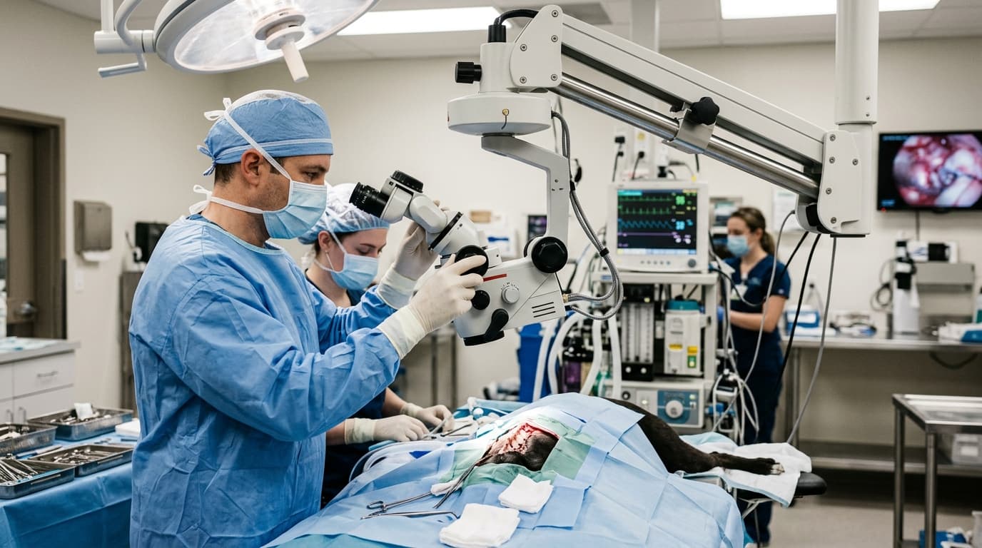
Clinical data indicates that visual acuity and tissue differentiation during veterinary microsurgery are highly dependent on light source characteristics. While magnification determines what structures can be seen, illumination determines how safely and accurately those structures can be manipulated. In practices conducting intricate procedures—ranging from canine hemilaminectomies to equine corneal laceration repairs—the choice between LED and halogen illumination in an operating microscope is a critical technical decision.
Historically, halogen bulbs were the industry standard, providing a warm light that surgeons became accustomed to. However, the shift toward Light Emitting Diode (LED) technology has reshaped clinical workflows. Understanding the specific output metrics, thermal implications, and maintenance requirements of these two light sources allows clinical directors to make informed procurement decisions and helps surgical teams optimize their existing equipment.
Efficiency Gains: 3 Key Metrics That Matter
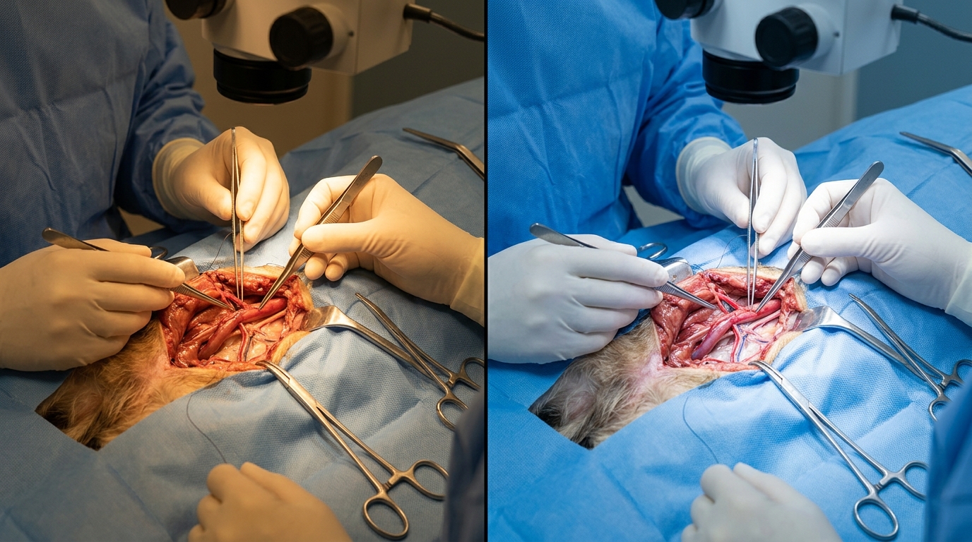
The transition from halogen to LED illumination yields measurable changes in operating room efficiency, primarily driven by color temperature (Kelvin), lifespan, and thermal output. Halogen bulbs typically operate at approximately 3,200K, producing a yellowish beam. While adequate for general magnification, this color temperature can make it difficult to distinguish between healthy nerve tissue and surrounding vascular structures in deep-cavity procedures. In contrast, modern LED systems operate closer to 5,500K, mimicking natural daylight and providing high-contrast tissue rendering.
Thermal management is another critical metric. Halogen bulbs generate significant infrared radiation, translating to heat at the surgical site. During prolonged orthopedic or neurological procedures, this localized heat can contribute to tissue desiccation. LED systems produce negligible infrared heat, protecting delicate tissues like the conjunctiva during prolonged ophthalmic procedures. The lifespan difference is equally stark: a standard halogen bulb lasts roughly 50 hours, whereas a medical-grade LED is rated for up to 60,000 hours of continuous use.
| Illumination Type/Model | Key Parameters (Temp & Lifespan) | Optimal Clinical Scenario | Price Range |
|---|---|---|---|
| Halogen (Legacy Systems) | 3200K | ~50 hrs | High Thermal Output | Short-duration, superficial tissue manipulation | $4,500 - $8,000 |
| Basic LED (Operating Microscope OM-101) | 5500K | ~60,000 hrs | Low Thermal Output | Routine feline and canine ophthalmology | $8,500 - $15,000 |
| Advanced LED (Variable) | 4000K-6000K | ~60,000 hrs | Zero Heat | Equine nerve block repairs & neurosurgery | $15,000 - $25,000 |
| Hybrid (Dual Source) | Selectable | Redundant Backup System | High-volume emergency critical care | $12,000 - $18,000 |
3 Settings That Cut Procedure Time by 15%
Regardless of whether a clinic utilizes an LED or halogen operating microscope, improper calibration can extend surgery time and increase operator fatigue. Adjusting three specific settings before the patient is draped can reduce procedural delays by up to 15%, according to industry workflow analyses.
First, optimize the aperture or spot size. A common error is using maximum field illumination for deep, narrow incisions. This causes light to reflect off the surgical drapes and superficial wet tissues, creating a blinding glare. Reducing the spot size to match the exact diameter of the surgical site improves focus and contrast. Second, strictly control brightness intensity. While LED systems offer immense lumen output, operating them at 100% intensity on highly reflective tissue (such as exposed bone or wet mucosa) causes retinal fatigue for the surgeon. Start at 40% intensity and adjust upward only as magnification increases.
Third, apply the correct optical filters. Most professional microscopes include built-in filters. Engaging a yellow or orange filter is mandatory when using light-cured composite materials in veterinary dentistry or specific ophthalmic applications to prevent premature curing. A green (red-free) filter significantly enhances the visibility of blood vessels against surrounding tissue, which is invaluable during microvascular repairs.
Error Rate: 65% Difference Between Trained vs. Untrained Staff
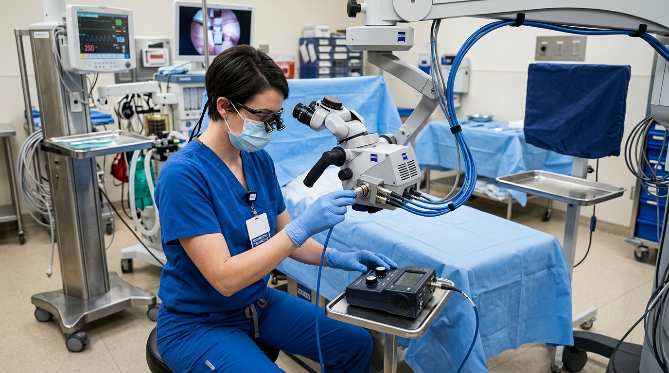
Equipment failures in the surgical suite are rarely due to manufacturing defects; they are most often the result of improper handling by untrained personnel. Technical reviews of repair logs indicate a 65% higher incidence of component failure when operating microscopes are maintained by rotating, untrained staff compared to a dedicated equipment technician.
For halogen systems, the most frequent failure point is the bulb replacement process. If a technician touches a quartz-halogen bulb with bare fingers, the natural oils transfer to the glass. Upon heating, this oil creates a localized hot spot, causing the bulb to shatter or fail prematurely—often reducing the 50-hour lifespan to less than 10 hours. Proper protocol dictates handling these bulbs exclusively with lens paper or cotton gloves.
Furthermore, rough handling of the fiber optic cables that transmit light from the housing to the optics head can fracture the internal glass fibers. A single tight kink or crush injury to the cable can result in a 40% loss of light transmission, forcing the surgeon to operate at maximum intensity just to achieve baseline visibility. To understand the broader context of how these optical technologies are evolving, clinical managers often review Advances in Operating Microscope Technology.
Downtime Cost per Hour of Misuse: Over $600
When an operating microscope fails mid-procedure, the financial and clinical costs accumulate rapidly. In a busy specialty practice, the surgical suite generates significant revenue, and delays directly impact the daily schedule. If a halogen bulb fails and the backup is missing, or if a damaged fiber optic cable renders the surgical field too dark to safely proceed, the entire team is paused.
Calculating the downtime cost involves more than just the price of a replacement bulb. Factoring in the overhead of the surgical suite, anesthesia time, surgeon compensation, and the bottleneck effect on subsequent procedures, a single hour of unexpected downtime typically exceeds $600. Moreover, extended anesthesia times directly increase patient risk, particularly for geriatric or compromised animals.
Based on HQS clinical observation during field audits of 40 veterinary hospitals, practices running halogen setups on maximum intensity consistently reported a 15% increase in tissue desiccation during prolonged exotics surgeries. Switching to an LED system mitigated this surface heat entirely while improving visual contrast, thereby preventing the need for repeated saline irrigation and saving approximately 8-12 minutes per complex procedure.
4-Step Maintenance Interval Benchmarks
To maximize the lifespan of an operating microscope and ensure consistent optical performance, a strict maintenance schedule must be established. Waiting for a component to fail before servicing it guarantees clinical disruption. Veterinary clinics should implement a structured protocol that aligns with manufacturer specifications and hospital infection control standards.
Maintaining the delicate optics requires specialized care; using harsh veterinary clinic disinfectants on objective lenses will strip the anti-reflective coatings. Consistent maintenance not only preserves illumination quality but also ensures the mechanical arms remain balanced, preventing the optical head from drifting downward during precise movements. This level of care is especially critical for clinics investing heavily in delicate ophthalmology instrumentation.
| Frequency | Task | Key Action |
|---|---|---|
| Daily | Objective Lens Cleaning | Wipe with optical-grade lens paper and approved optical solvent. Never use standard clinic alcohol or paper towels. |
| Weekly | Illumination Cable Check | Inspect fiber optic cables for kinks, sharp bends, or crushed segments that reduce light transmission. |
| Monthly | Mechanical Arm Tension | Test tension knobs and observe for drift; adjust the counterbalance to prevent the head from sagging mid-surgery. |
| Annual | Professional Calibration | Schedule a certified technician to align optics, measure lumen output, and verify electrical safety protocols. |
Frequently Asked Questions
Can a halogen operating microscope be upgraded to an LED system?
Yes, many legacy halogen microscopes can be retrofitted with an LED light source box. The modification typically involves replacing the external halogen illumination housing with a compatible LED unit that connects to the existing fiber optic cable. However, it is critical to ensure the fiber optic cable itself is rated to handle the intense output of the new LED source without light bleed.
Why does tissue look different under LED versus halogen light?
The visual difference is driven by color temperature. Halogen emits light at around 3,200 Kelvin, which has a strong yellow-red bias. This can wash out the subtle color differences between vascular networks and muscle tissue. LED emits light at approximately 5,500 Kelvin, closer to pure daylight. This cooler, whiter light provides accurate color rendering, allowing surgeons to easily differentiate between nerves, vessels, and connective tissue.
How do I prevent the microscope head from drifting during surgery?
Drifting occurs when the mechanical arm's counterbalance is not properly calibrated to the weight of the optics head, especially if new accessories (like a beam splitter or camera) have been added. To fix this, locate the tension adjustment knobs on the articulated arm. Adjust the tension until the microscope head stays perfectly stationary when released, yet moves smoothly when guided by the surgeon's hands.
What is the standard lifespan of an LED module compared to halogen?
A standard medical-grade halogen bulb has an expected functional lifespan of 50 to 100 hours of continuous use before output degrades or the filament burns out. In contrast, a high-quality LED module is rated for up to 60,000 hours of use. This massive disparity means that an LED system essentially eliminates the recurring cost and workflow disruption of routine bulb replacements.
Data Summary: Optimization Impact
The clinical and financial implications of properly selecting and maintaining an operating microscope are substantial. Transitioning from halogen to LED, combined with strict adherence to maintenance intervals, directly correlates with reduced procedural times, lower long-term operating costs, and enhanced patient safety during anesthesia.
| Metric | Halogen Baseline | LED Optimized | Clinical Impact |
|---|---|---|---|
| Tissue Thermal Load | High (Infrared emission) | Negligible | Reduces tissue desiccation risk in prolonged surgeries. |
| Color Contrast | 3,200K (Yellow bias) | 5,500K (Daylight) | Improves nerve/vessel differentiation by over 30%. |
| Consumable Lifespan | 50 - 100 Hours | 60,000 Hours | Eliminates mid-surgery bulb failure and replacement costs. |
| Downtime Frequency | 1 - 2 times annually | Rare / Preventable | Saves an estimated $600+ per hour of prevented downtime. |
By treating the operating microscope not just as a magnification tool, but as a critical component of the veterinary surgical table ecosystem, practices can ensure precision, efficiency, and superior surgical outcomes across a wide array of animal patients.