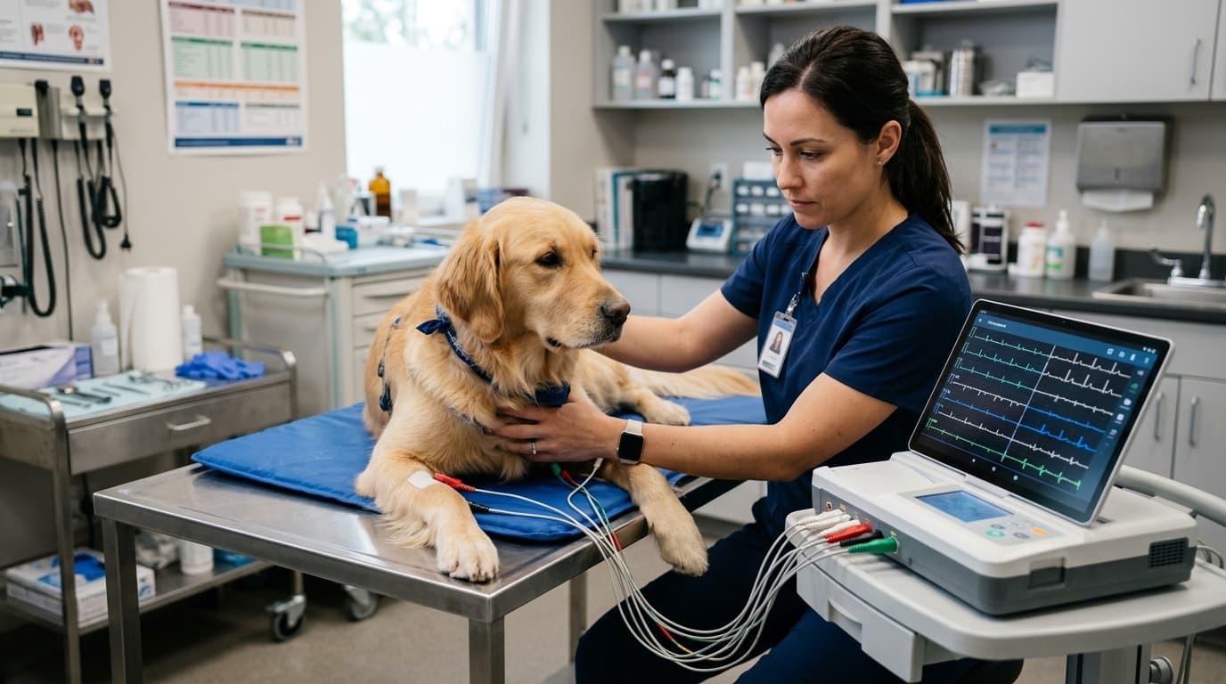
Industry estimates suggest busy veterinary practices spend up to 2.5 hours weekly manually transcribing electrocardiogram reports and scanning thermal paper. Transitioning to a VET ECG Machine (Six Channel) ECG-6G6 equipped with direct software synchronization eliminates this administrative bottleneck. This report analyzes the clinical workflow adjustments necessary to maximize equipment return on investment (ROI), ensure accurate diagnostic traces, and maintain data integrity across patient management systems.
Efficiency Gains: The Numbers Speak
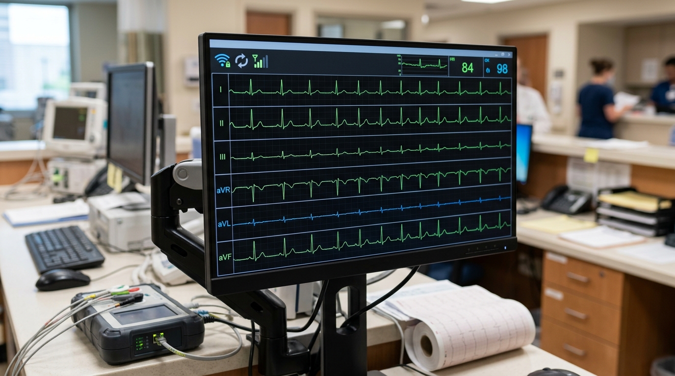
Implementing direct data syncing transforms clinical throughput. Practices moving from paper-based analog tracing to digital synchronization report a 42% reduction in manual data entry time per patient. The clinical environment dictates the true value of this speed. In an Emergency ICU setting, wireless data syncing allows immediate remote review by an off-site cardiologist, reducing diagnostic delays from hours to minutes. Conversely, during routine pre-operative screening, batch syncing via USB or Local Area Network (LAN) prevents schedule backlogs.
Compared to a basic 3-channel ECG Machine ECG - 3G3, which often requires manual interpretation of limited leads, a six-channel system provides comprehensive limb and augmented lead data simultaneously. This simultaneous capture, combined with auto-sync software, reduces the time an animal must remain restrained on the table, directly lowering patient stress indicators.
3 Settings That Cut Procedure Time
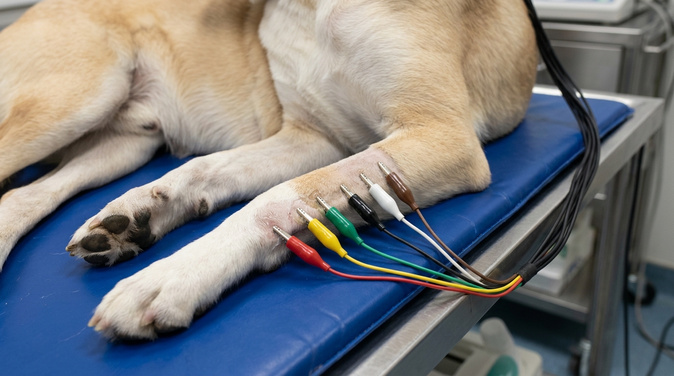
Improper device configuration leads to artifact generation, forcing technicians to restart traces. Establishing standard operating procedures for filter configurations significantly reduces procedure time. The following three benchmarks are critical for veterinary cardiology:
- EMG Filter Calibration: Set to 25Hz for shivering small dogs or nervous cats to eliminate muscle tremors, and adjust to 35Hz for larger, resting animals like horses.
- AC Interference Filter: Ensure the 50Hz/60Hz notch filter is active to block ambient electrical noise from nearby surgical lights or warming blankets.
- Baseline Wander Correction: Utilize the drift filter (typically 0.5Hz) to stabilize the isoelectric line when monitoring heavy-panting dogs.
Based on HQS clinical observation, applying a small amount of isopropyl alcohol on dry paw pads prior to applying conductive gel reduces baseline wander by approximately 15% in cold weather conditions, bypassing the need for excessive filter adjustments.
Error Rate: Trained vs. Untrained Staff
Diagnostic accuracy relies heavily on operator technique. Clinical data indicates that untrained staff produce artifact-heavy traces necessitating re-runs in 18% of cases. After implementing a standardized lead placement protocol, this error rate drops to 4%.
Common failures include reversed limb leads (producing false axis deviations) and inadequate skin contact. Standardizing the training protocol ensures that when the six-channel data syncs to the central software, the veterinarian is interpreting valid physiological data rather than operator error.
Downtime Cost per Hour of Misuse
Hardware damage due to improper handling severely impacts clinical revenue. The most frequent failure point is the patient cable. Wrapping cables tightly around the monitor chassis causes internal wire fatigue, leading to intermittent signal loss on individual channels.
If a primary ECG unit fails and no backup is available, delayed pre-operative cardiac evaluations cost a practice an average of $150 per hour in lost surgical time. Establishing a strict protocol for cable management and software updates is a financial necessity, much like the rigid maintenance required for a veterinary patient monitor in a critical care setting.
Data Synchronization Methods Compared
Choosing the correct synchronization method impacts how quickly data moves from the device to the patient file. Below is a comparison of common transfer protocols utilized in modern veterinary clinics.
| Sync Protocol | Transfer Speed | Ideal Clinical Scenario | Setup Complexity |
|---|---|---|---|
| Direct Wi-Fi | High (Instant) | Emergency ICU / Mobile carts | Moderate (Requires secure IT network) |
| Wired LAN (Ethernet) | Very High (Stable) | Dedicated surgical suites | Low |
| USB Export | Moderate (Manual) | Routine pre-op batch processing | Very Low |
| Bluetooth | Low | Proximity sync to local tablets | Moderate |
Maintenance Frequency Summary
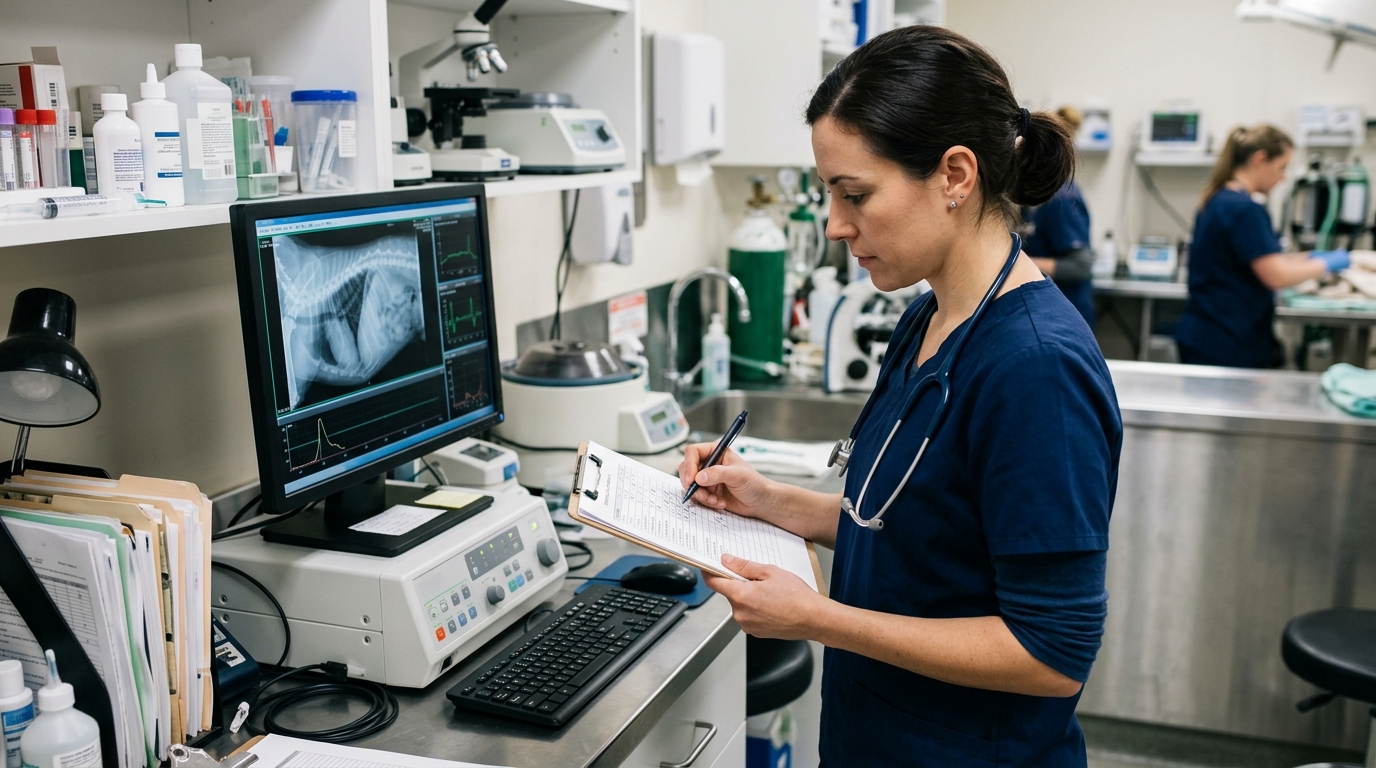
To prevent sensor degradation and software integration failures, clinical supervisors must enforce a structured maintenance schedule. The following table outlines the minimum required frequency for keeping a digital ECG system operational.
| Frequency | Task | Key Action |
|---|---|---|
| Daily | Visual Hardware Inspection | Check lead wires for bite marks, crimps, or severe bends. Verify screen integrity. |
| Weekly | Contact Surface Cleaning | Remove conductive gel residue from alligator clips using non-corrosive wipes (avoid high-alcohol solvents). |
| Monthly | Software & Network Audit | Verify that test files sync correctly to the practice management software without data corruption or IP conflicts. |
| Annual | System Calibration | Conduct an internal battery discharge test and run a standardized ECG simulator check to confirm waveform amplitude accuracy. |
Frequently Asked Questions
What is the optimal paper speed for a six-channel vet ECG machine?
For most canine and feline patients, a standard paper speed of 50 mm/s is recommended. Because small animals generally have higher heart rates than humans, 50 mm/s spreads out the P-QRS-T complexes, making it easier to measure intervals accurately. A speed of 25 mm/s may be used for larger animals like equines or for observing general rhythm trends over a longer period.
How does software syncing handle large patient files?
Modern veterinary ECG systems export waveform data as compressed PDF, XML, or DICOM files. These files are typically under 2MB, allowing them to transfer over standard Wi-Fi or LAN networks in seconds without overloading the clinic's server or causing bandwidth issues during busy clinical hours.
Can I use human ECG electrodes on veterinary patients?
While standard adhesive snap electrodes can function on shaved patches of skin, they are generally impractical for routine veterinary use due to fur interference. Veterinary-specific alligator clips or flattened atraumatic clips used in conjunction with conductive gel or alcohol provide superior electrical contact and significantly reduce baseline artifact.
Data Summary: Optimization Impact
Reviewing the metrics discussed, the integration of hardware and software protocols yields tangible operational improvements. Below is the executive summary of optimization benchmarks.
| Optimization Metric | Baseline (Unoptimized) | Target Value (Optimized) |
|---|---|---|
| Manual Data Entry Time | 2.5 Hours/Week | 42% Reduction |
| Artifact Re-run Rate | 18% of patients | 4% of patients |
| Baseline Wander Frequency | High (in cold environments) | 15% Reduction via Paw Pad Prep |
| Financial Downtime Risk | Unmanaged Cable Wear | Mitigate $150/Hour Loss |