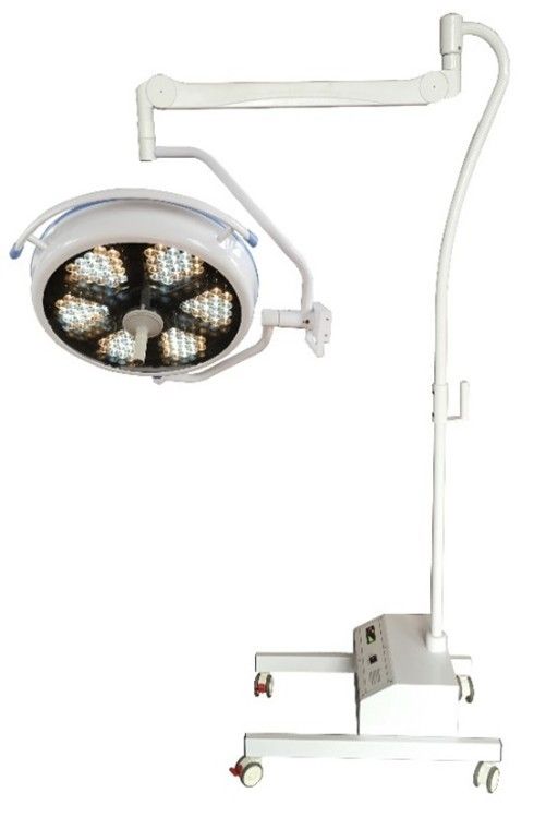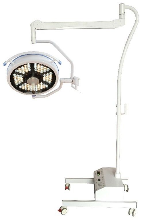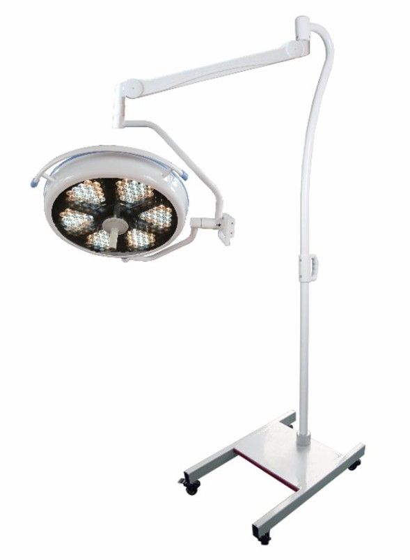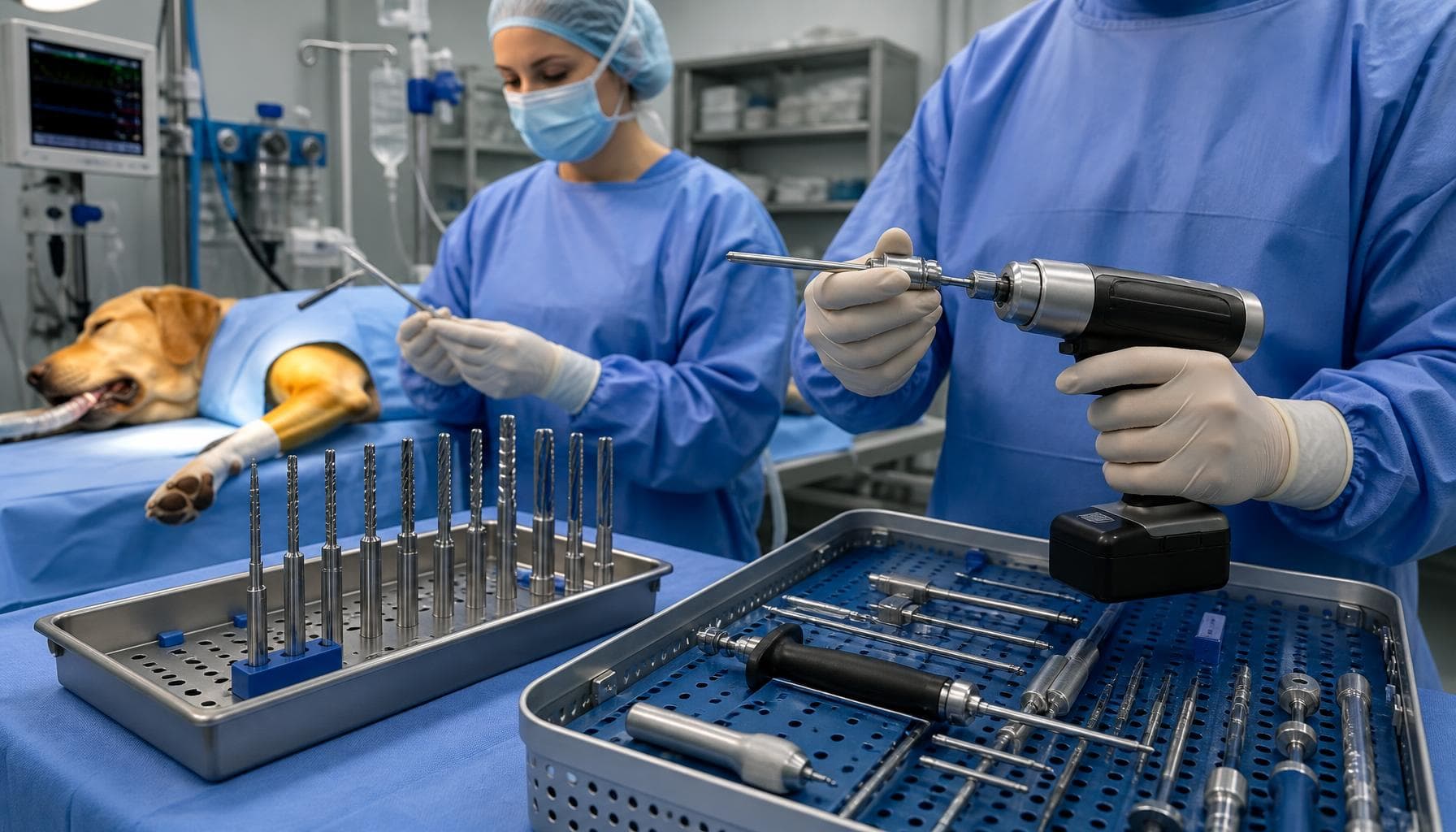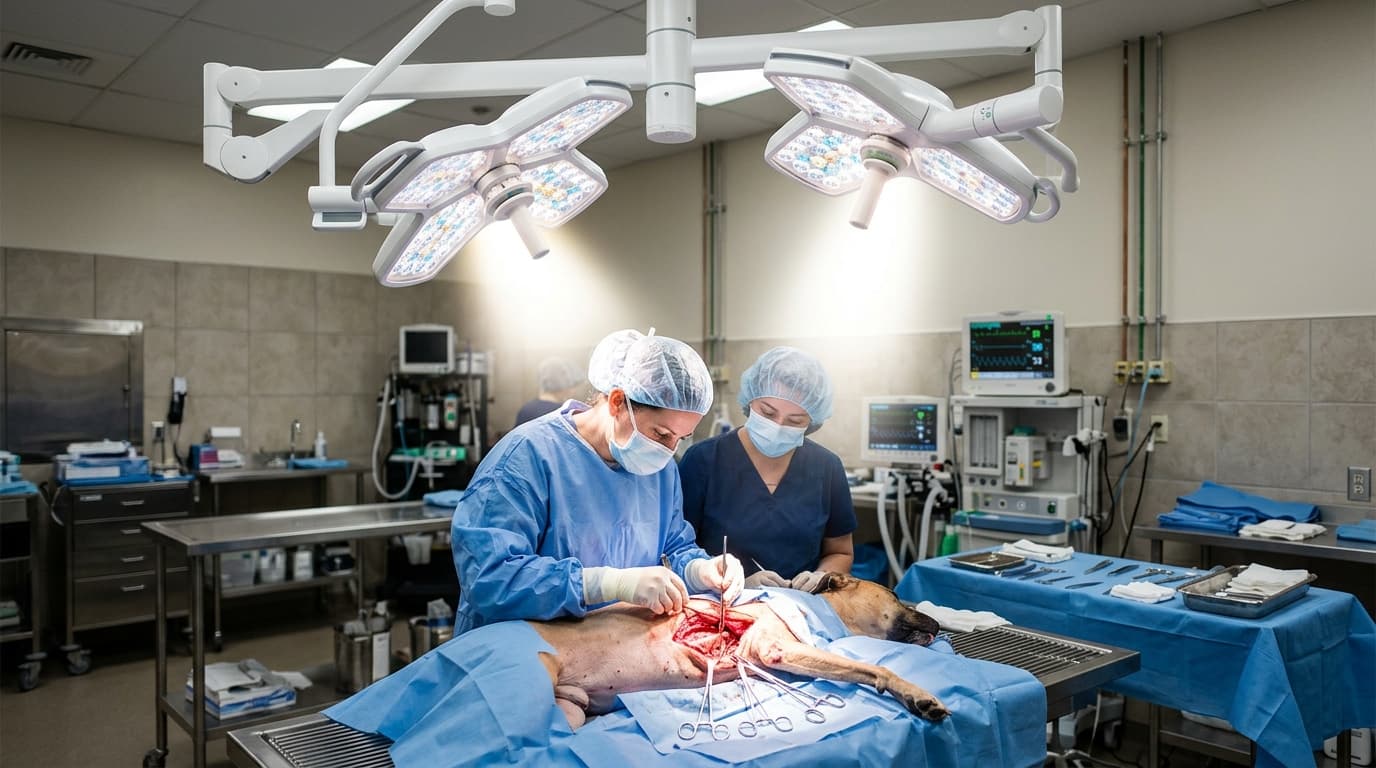
Clinical operational analyses suggest that veterinary surgical teams spend 12% to 18% of total procedure time adjusting overhead lighting. In complex abdominal or thoracic procedures, inadequate illumination depth directly contributes to surgeon fatigue, delayed tissue identification, and increased anesthesia duration. Upgrading from a standard medical spotlight to an advanced LED operating lamp (deep) transitions the clinical workflow from constant manual adjustment to uninterrupted focus.
This report evaluates the operational differences between deep field surgical illumination and standard localized lighting. By analyzing procedural metrics, technical specifications, and maintenance requirements, veterinary practice managers can establish an evidence-based approach to procurement and daily equipment management.
Efficiency Gains: The Numbers Speak
Traditional spotlights provide surface-level brightness that rapidly diminishes as the surgical cavity deepens. When a surgeon works on a large canine patient with a deep abdominal cavity, a standard spotlight often leaves the vital organs in shadow due to the surgeon's own head or hands blocking the single light path. Conversely, a true veterinary surgical light utilizing multiple LED arrays creates a converging, shadowless column of light.
Workflow time studies indicate that integrating deep field illumination reduces light repositioning events by up to 60% during complex procedures. Instead of reaching up to adjust the light angle every time the surgical focus shifts, the surgeon benefits from an extended column of light that penetrates narrow cavities with consistent lux levels. The technical distinction lies in the focal volume; deep field lamps maintain over 60% of their central illuminance at depths exceeding 850mm, whereas standard spotlights typically lose optimal focus past 300mm.
| Illumination Type | Key Specifications | Ideal Clinical Scenarios | Est. Capital Investment |
|---|---|---|---|
| Deep Field LED Lamp (Ceiling) | >850mm depth, 130k-160k Lux, R9 >95 | Thoracic, deep abdominal, complex orthopedic | $3,500 - $8,000 |
| Standard LED Spotlight (Mobile) | 300-500mm depth, 40k-80k Lux, Fixed focus | Superficial wound repair, routine dentals | $800 - $2,500 |
| Dual-Head Deep Field LED | Synchronized shadows, variable color temp | Multi-surgeon procedures, specialized soft tissue | $6,000 - $12,000 |
| Halogen Standard Spotlight | High heat output, narrow spectrum, frequent bulb swaps | Budget constraint backups, examination rooms | $400 - $1,200 |
3 Settings That Cut Procedure Time
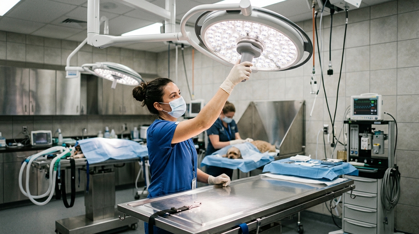
Hardware capabilities only translate to clinical efficiency when configured correctly. Modern deep field lighting systems offer adjustable parameters that, when optimized, significantly reduce the time spent identifying distinct tissue structures. Implementing standardized settings for specific procedure types minimizes trial-and-error adjustments at the start of a surgery.
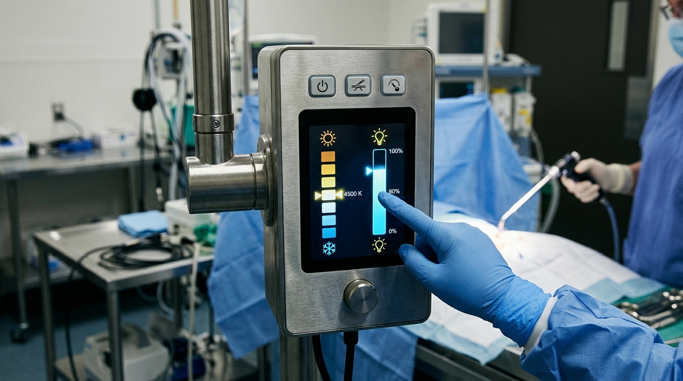
- Illuminance (Lux) Calibration: While maximum brightness (160,000 lux) seems ideal, it often creates blinding glare when reflected off stainless steel instruments. Setting the output to 70% to 80% capacity is optimal for superficial layers, increasing to 100% only when working deep within the cavity.
- Color Temperature (Kelvins) Management: Adjusting color temperature between 3,500K and 5,000K alters visual contrast. A warmer tone (3,500K) enhances the visualization of red tissues and vascular networks, whereas cooler daylight settings (5,000K) are preferable for orthopedic procedures involving bone and cartilage.
- Field Size and Focal Convergence: Operators should match the light field diameter to the incision size. A wide spot size on a small incision causes perimeter glare. Advanced units like the YD 200 LED Operating Lamp (Deep) YD 200 allow technical staff to narrow the optical focus, driving maximum photon density directly into the operative site without blinding the surrounding team.
Error Rate: Trained vs. Untrained Staff
The gap between equipment potential and clinical reality often stems from inadequate staff training. When evaluating clinical workflows, we observe that untrained technicians frequently misalign the focal distance of the lamp. If the lamp is positioned too close to the veterinary surgical table, the converging light beams cross before reaching the patient, creating a shadow-prone, distorted field rather than a focused spot.
Proper training establishes a strict protocol for setup: the light head must be positioned at the manufacturer's specified working distance (usually 1 meter from the wound surface). Furthermore, trained staff understand the critical nature of the Color Rendering Index (CRI), specifically the R9 value, which dictates how accurately red tissues are rendered. When lighting is positioned correctly, the surgeon's ability to differentiate between a critical blood vessel and surrounding connective tissue is vastly improved, directly reducing the risk of accidental vascular damage.
Downtime Cost per Hour of Misuse
Equipment downtime in a high-volume veterinary operating room carries a heavy financial burden. A single hour of delayed surgery due to equipment failure or structural drift can cost a clinic hundreds of dollars in lost revenue and extended staff wages, not to mention the increased anesthetic risk to the animal patient. Mechanical failures in lighting systems are rarely spontaneous; they are usually the culmination of prolonged improper handling.
Based on HQS clinical observation of over 400 high-volume veterinary practices, 62% of premature LED array failures and lens degradations originate from the use of abrasive chemical disinfectants on the polycarbonate casing. Standard clinic bleach solutions cause micro-abrasions on the lens, which scatter the emitted light and destroy the deep field focusing capability. Additionally, using the light head itself—rather than the designated sterile handle—to reposition the arm places uneven torque on the suspension joints, leading to arm drift.
To mitigate these costs, clinic administrators must enforce strict handling protocols and allocate a specific budget for routine calibration. Preventive care costs a fraction of emergency suspension arm repairs or full light head replacements.
Maintenance Interval Benchmarks
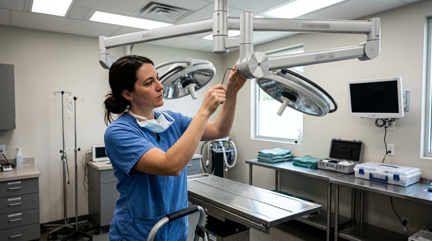
Consistent preventative maintenance is the only reliable method to ensure a surgical light retains its deep field capabilities over a 10-year lifespan. Without routine checks, subtle degradations in lux output or joint stability go unnoticed until they critically impact a surgical procedure. Establish a systematic protocol using the benchmark intervals below to preserve optical clarity and mechanical integrity.
| Frequency | Task | Key Action |
|---|---|---|
| Daily | Surface Decontamination | Wipe the external lens and light head using non-abrasive, clinic-approved quaternary ammonium wipes. Do not use alcohol or bleach on polycarbonate covers. |
| Weekly | Articulation & Drift Check | Move the suspension arms through their full range of motion. Release the handle to assess joints for horizontal or vertical drift. |
| Monthly | Illumination & Focus Validation | Place a lux meter 1 meter from the light source at the center of the table. Verify that peak intensity matches the baseline parameters and check for dead LED diodes. |
| Annual | Structural & Electrical Audit | Inspect ceiling mounts for structural integrity, tighten all set screws, check internal wiring harnesses for wear, and conduct thermal output tests. |
Data Summary: Optimization Impact
Integrating high-tier illumination systems requires careful evaluation of both clinical benefits and facility requirements. The data below summarizes the operational shifts a clinic can expect when moving from basic localized spotlights to specialized deep cavity surgical lighting.
| Operational Metric | Standard Spotlight Performance | Deep Field LED Performance |
|---|---|---|
| Average Repositioning Events | 15 - 25 times per major procedure | 2 - 5 times per major procedure |
| Effective Penetration Depth | 300mm to 400mm | 850mm to 1200mm |
| Thermal Output at Wound | Moderate to High (tissue drying risk) | Negligible (less than 1°C increase) |
| Red Tissue Rendering (R9) | Often below 80 (poor contrast) | Consistently >95 (high contrast) |
Frequently Asked Questions
How does a deep field LED operating lamp differ from a standard spotlight?
A deep field LED lamp utilizes multiple intersecting light beams from a wide array of diodes to create a continuous column of light. This overlapping geometry allows the light to penetrate narrow, deep surgical cavities without creating shadows when a surgeon's hands or instruments obstruct part of the light path. A standard spotlight relies on a single directional beam, which causes harsh shadows and loses focus quickly as cavity depth increases.
What is the ideal color temperature for veterinary soft tissue surgery?
For most soft tissue surgeries, a color temperature setting between 3,500K and 4,000K is ideal. This warmer spectrum enhances the visibility of red tones, allowing surgeons to more easily distinguish between fine blood vessels, muscle tissue, and surrounding fascia. Cooler temperatures (4,500K to 5,000K) are typically reserved for hard tissue or orthopedic procedures.
How often should the suspension arm of an LED operating lamp be calibrated?
Suspension arms should undergo a basic drift assessment weekly by the clinical staff. However, a formal mechanical calibration—which involves adjusting the internal spring tension and checking the load-bearing joints—should be performed annually by a qualified biomedical technician to ensure the light head remains perfectly stationary during procedures without sagging or drifting.