sales@hqs-vetcare.com
86-19805651775
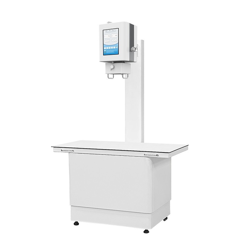
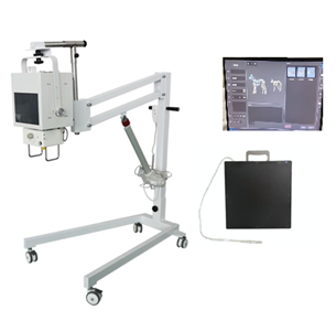
DR-A10 Veterinary Digital
Radiography System
Digital radiography offers a significant advancement in image acquisition compared to traditional film-based radiography. In conventional methods, after capturing X-ray images on film, a time-consuming development process is required to reveal the diagnostic results. However, with digital radiography, this bottleneck is eliminated. The technology enables near-instantaneous acquisition of high-quality images, providing veterinarians with immediate access to diagnostic information.
The process involves the use of digital sensors to capture X-ray images, which are then promptly available on a computer or viewing console. This rapid availability is especially crucial in urgent or emergency cases where quick assessments and timely decision-making are paramount. Veterinarians can efficiently analyze the digital images, allowing for prompt diagnosis and the formulation of necessary treatment plans. The elimination of the development step not only accelerates the diagnostic process but also contributes to more efficient patient care, ensuring that interventions can be initiated promptly based on the real-time information provided by digital radiography.
Features
1. Applicable to multiple scenarios, cost-effective choice.
2. Removable, integrated integrated X-ray head, suitable for various use scenarios such as hospital and field.
3. Three exposure modes: remote control, handle control and touch screen control, to meet the needs of different scenes.
4. 17*17 inch amorphous silicon flat panel detector, clear imaging and clear layers.
5. AED automatic exposure technology to ensure image quality, reduce scrap rate, and reduce repeated shooting.
6. Bizcon image acquisition software with independent intellectual property rights, simple and intuitive workflow design, and practical measurement tools for medical specialists.
Technical Specifications
PARAMETER | SPECIFICATION | |
Fixed SID distance adjusted to 1000 mm | ||
Table Top Size | 1210 mm x 610 mm | |
Whole Machine Dimension | 1200 mm x 860 mm x 2000 mm | |
PARAMETER | a-Si TFT Detector | |
Pixel Size | 139μm | |
Pixel Matrix | 2912 x 2912 | |
AD Conversion | 16 bits | |
Spatial Resolution | 3.6 lp/mm | |
Imaging Area | 405mm × 405mm (17” x 17”) | |
Dimension | 460mm x 460mm x 15mm | |
Weight | 4Kg | |
PARAMETER | Portable X-ray Machine | |
High Frequency | 40 KHz | |
Single Phase | 220VAC+-10%, 5 KW | |
Output Power | 100mA and 120Kvp | |
X-ray tube H.U. continual monitoring for X-ray tube protection | ||
Kvp Radiographic range | from 40 to 120 Kvp | |
mA Radiographic range | from 10 to 100 mA | |
Anatomical program APR (Kvp, mAs, AEC, focal spot, etc) | ||
Light and sound indication for X-ray exposure | ||
Hand-Switch for preparation and exposure control. | ||
Manual collimator with 6 pair of leafs. | ||
Accessory rails (cones, filters, etc.) | ||
Field LED lamp of 160 LUX | ||
Light indicator for alignment with Bucky. | ||
Retractable measuring tape. | ||
Light indicator for alignment with Bucky. | ||
Retractable measuring tape | ||
PARAMETER | Workstation and S/W (B-Console vet) | |
Preview & Post processing Time : 3 sec | ||
KV, mA, mAS, Thumb nail View, patient Info and so on | ||
Main Screen Console: Veterinary patient information | ||
Automatic Pre & Post image enhancement. | ||
Expands image processing for optimized viewing | ||
Enhances variable density and form shading | ||
Variety Measurements | ||
Application measurements & teaching tools (such as VHS, HD, TPLO, TTA etc) | ||
One key to capture images into Clipboard | ||
DICOM sending and Report writing and Printing | ||
Easy Calibration & User setting | ||
Contact: Mr.Shao
Phone: 86-19805651775
E-mail: sales@hqs-vetcare.com
Whatsapp:86-19805651775 / 81-08068730425
Add: Room 402, Unit 6, Building 27, Pingchou District 2, Jiangdong Street, Yiwu, Jinhua, Zhejiang Province
WhatsApp Chat