sales@hqs-vetcare.com / info@hqs-vetcare.com
86-19805651775
Veterinary medicine and technologies are growing and changing everyday, and with these changes medical equipment is modernizing and expanding. Change and expansion produce updated and innovative medical equipment which make veterinary services and practices more convenient and useful for doctors, patients, and pet owners. Keeping up with these changes and new advances can help to add value to your practice. Going one step further, these new methods of imaging can be used for thorough dental x-rays on numerous animals. Using digital veterinary equipment not only advances your practice, it also ensures the safest and most up-to-date care for the animals. At the end of the day, conveniently and thoroughly meeting the needs of the patients is key to any medical practice.
Features
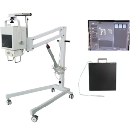
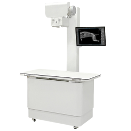
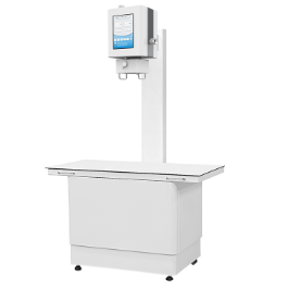
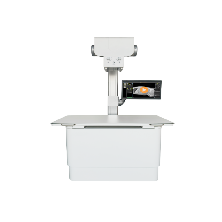
V-DRF30 developed specifically with veterinarians and their work flows in mind, is a combination of mobile DR system and C-arm system(2-in-1), enabling the veterinarians to enjoy powerful output in X-ray room and conveniences diagnosis in operating room.
DRF can switching between digital X-ray and live X-ray Video fruoroscopy mode without any adjustment to your patient, moving between X-ray room and operating room freely.
Plenty of new Applications with live X-ray video fluoroscopy beyond digital X-ray.
-Evaluating urinary issues
-Studies of soft tissue or ligament damage
-Finding small fractures
-Diagnosing tracheal collapse
-Investigating possible jaw or skull fractures
-Diagnosing a hernia
-Orthopedical issues
-Swallowing/Esophageal studies
- Dual monitors: 19”touch monitor + 24” main monitor
Technical Specifications
PARAMETER | a-Si TFT Dynamic Detector | |
Pixel Size | 139μm | |
Pixel Matrix | 3072 x 3072 | |
AD Conversion | 16 bits | |
Spatial Resolution | 3.6 lp/mm | |
Imaging Area | 405mm × 405mm (17” x 17”) | |
Dimension | 460mm x 460mm x 15mm | |
Weight | 4Kg | |
PARAMETER | X-ray Generator Features | |
High Frequency | 250 KHz | |
Single Phase | 220/240VAC 32 KW | |
Output Power | 400 mA and 150Kv | |
X-ray tube H.U. continual monitoring for X-ray tube protection | ||
Kvp Radiographic range | from 40 to 150 Kvp | |
mA Radiographic range | from 10 to 400 mA | |
Anatomical program APR (Kvp, mAs, AEC, focal spot, etc) | ||
Dual Switch (Foot and Hand-Switch) for preparation and exposure | ||
PARAMETER | X-ray Tube | |
Canon(Toshiba) | E7239X | |
Maximum Tension | 125 kV | |
Focus sizes | Small focus 1.0 mm, Large focus 2.0 mm | |
Anode target angle : | 14 º | |
Anode Rotation | 2700 | |
PARAMETER | Manual Collimator | |
Manual collimator with 6 pair of leafs | ||
Accessory rails (cones, | ||
Field LED lamp of 160 LUX | ||
Light indicator for alignment with Bucky | ||
Retractable measuring tape | ||
PARAMETER | Workstation and S/W (B-Console vet) | |
Preview & Post processing Time : 3 sec | ||
KV, mA, mAS, Thumb nail View, patient Info and so on | ||
Main Screen Console: Veterinary patient information | ||
Automatic Pre & Post image enhancement. | ||
Expands image processing for optimized viewing | ||
Enhances variable density and form shading | ||
Variety Measurements | ||
Application measurements & teaching tools (such as VHS, HD, TPLO, TTA etc) | ||
One key to capture images into Clipboard | ||
DICOM sending and Report writing and Printing | ||
Easy Calibration & User setting | ||
Contact: Mr.Shao
Phone: 86-19805651775
E-mail: sales@hqs-vetcare.com / info@hqs-vetcare.com
Whatsapp:86-19805651775 / 81-08068730425
Add: Room 402, Unit 6, Building 27, Pingchou District 2, Jiangdong Street, Yiwu, Jinhua, Zhejiang Province
WhatsApp Chat