Thermal necrosis remains a primary cause of delayed union, aseptic loosening, and implant failure in veterinary orthopedics. Bone tissue exposed to temperatures exceeding 47°C for just 60 seconds undergoes irreversible cellular death. In veterinary clinical practice, mitigating this risk requires strict control over instrument sharpness, applied feed pressure, and rotational speed. Industry observation suggests that standardizing equipment maintenance and monitoring instrument wear can cut thermal necrosis risk by up to 62%, drastically improving postoperative recovery metrics.
This report analyzes the mechanical parameters that dictate safe orthopedic preparation, providing data-driven baselines for technical staff and surgeons. By treating bit sharpness and thermal management as measurable variables rather than subjective preferences, veterinary hospitals can improve procedure times, reduce hardware failure, and extend the lifespan of their surgical capital equipment.
Thermal Necrosis: The 47°C Threshold Data
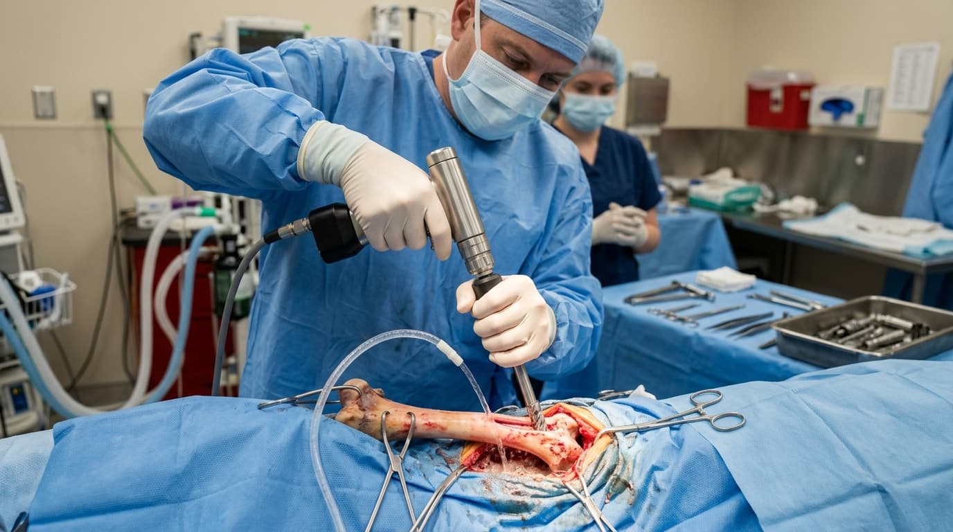
Heat generation during intramedullary reaming or plating preparation is a function of friction and cutting efficiency. When a dull bit meets dense cortical bone, such as in a canine tibial plateau leveling osteotomy (TPLO), the mechanical energy converts directly into thermal energy rather than shearing away bone tissue. Healthy cancellous bone is slightly more forgiving, but cortical bone lacks the vascularity to dissipate rapid heat spikes.
Maintaining temperatures below the critical 47°C threshold requires a combination of sharp cutting flutes, appropriate torque, and consistent irrigation. When a bit loses its primary cutting edge, surgeons instinctively apply higher axial pressure. This increased feed pressure forces the blunt instrument against the bone, rapidly elevating the local temperature to 50°C or higher within seconds, leading to localized osteonecrosis and compromising the implant-bone interface.
3 Reamer Drill Settings That Cut Procedure Time
Optimizing equipment settings is as critical as maintaining bit sharpness. Operating a veterinary bone drill and saw system outside of its intended parameters accelerates wear and increases heat transfer. To safely minimize procedure time, hospitals must standardise three specific operational metrics:
- Rotational Speed (RPM): Reaming requires high torque and low speed. Operating between 150 and 300 RPM allows the cutting flutes to engage and remove bone effectively without generating excessive friction.
- Irrigation Volume: Continuous room-temperature sterile saline irrigation at a minimum flow rate of 30–50 mL/minute is required to flush debris from the flutes and act as a thermal sink.
- Pecking Rhythm: Instead of continuous axial pressure, applying a 2-second engagement followed by a 1-second retraction clears bone swarf, dropping temperatures by an average of 4°C per cycle compared to continuous plunging.
Bit Wear Metrics: 5 Indicators for Replacement
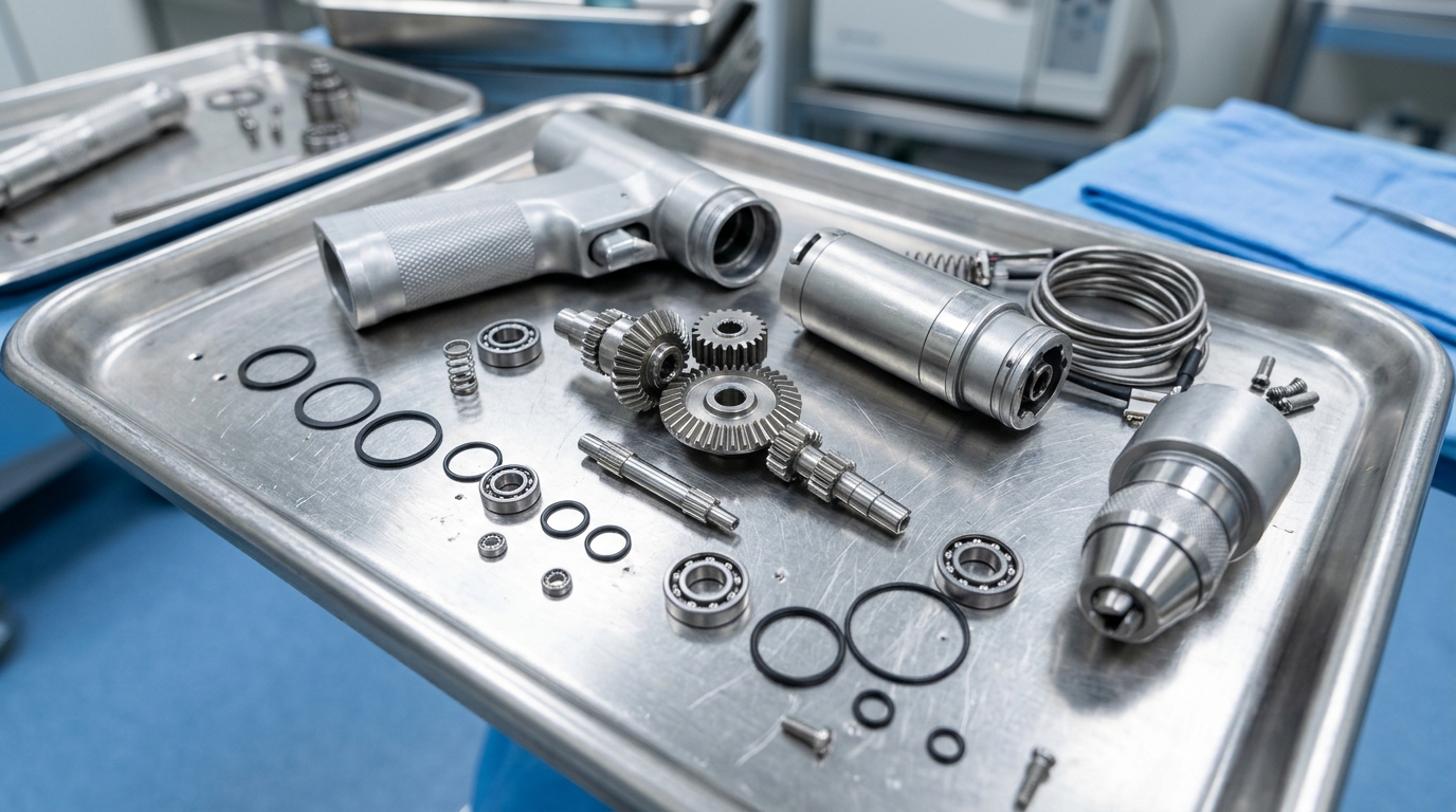
Visual inspection often fails to identify micro-fractures or dulling on cutting edges. Utilizing the Reamer Drill RD - 4011 or similar high-torque systems requires a strict replacement protocol for consumables. Staff should assess the following physical indicators to determine bit viability:
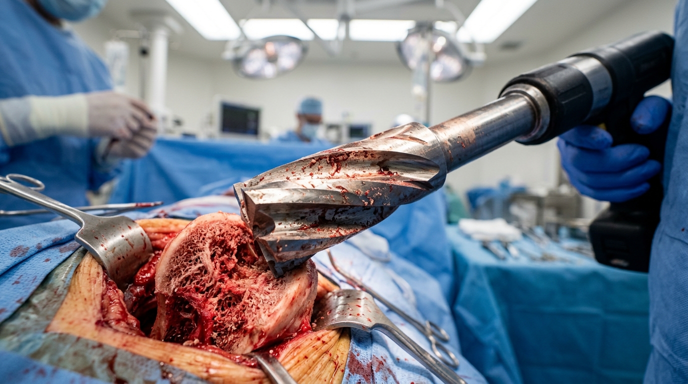
- Flute Galling: The accumulation of fused bone material or metallic smearing on the cutting flutes, indicating excessive heat during prior use.
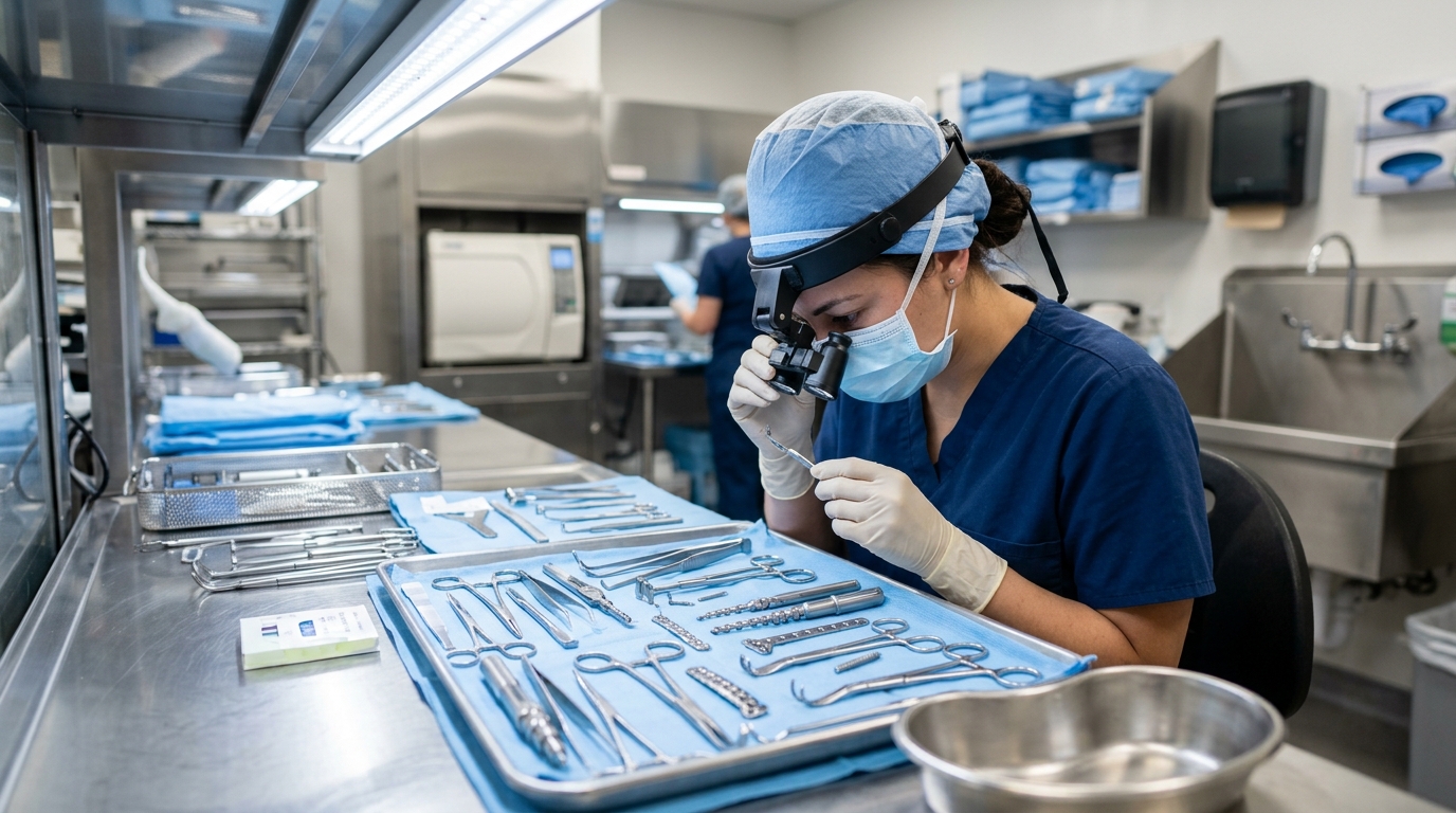
- Edge Rounding: Loss of the sharp, distinct primary cutting edge, observable under 3x to 5x magnification during sterile processing.
- Discoloration: Blue or straw-colored tempering marks on the stainless steel, which confirm the bit has exceeded safe operating temperatures and lost its structural hardness.
- Chatter Marks: Asymmetrical wear patterns that cause the bit to vibrate or "walk" off the target insertion point before engaging the cortex.
- Cycle Count: Tracking the number of sterilization cycles. Industry benchmarks recommend assessing heavy-use orthopedic bits for replacement after 20 to 30 clinical cycles.
Error Rate: Trained vs. Untrained Staff
Based on HQS clinical observation across multiple veterinary orthopedic centers, untrained technical staff often mistake a lack of cutting progress for a lack of motor power, prompting them to increase RPMs instead of replacing a dull bit. This operational error compounds the thermal necrosis risk.
When staff are trained to recognize the tactile feedback of a sharp bit—which pulls itself into the medullary canal with minimal axial force—they intervene earlier. Training programs that establish strict "feel and listen" protocols result in a measurable decrease in surgical complications. A sharp bit produces consistent bone chips, whereas a dull bit produces fine bone paste and smoke, an immediate indicator that the thermal threshold has been breached and necrosis is actively occurring.
Downtime Cost per Hour of Misuse
Applying excessive feed pressure on a dull bit does not merely damage the patient's bone; it transfers massive axial load back into the equipment's internal gearing and motor housing. Veterinary clinics frequently underestimate the financial impact of this mechanical abuse.
Operating a high-torque drill with blunt consumables draws excess electrical current, overheating the handpiece and deteriorating the internal seals. This practice leads to premature motor failure and autoclave-induced moisture damage once the seals are compromised. The resulting equipment downtime disrupts surgical schedules, delays critical patient care, and incurs significant repair costs. Establishing clear protocols for veterinary reamer drill clinical best practices is a direct investment in the hospital's financial stability and equipment longevity.
Maintenance Interval Benchmarks
To prevent performance degradation and ensure clinical safety, veterinary practices must adopt a rigorous maintenance schedule. The following protocol outlines the baseline checks necessary for high-demand orthopedic equipment.
| Frequency | Task | Key Action |
|---|---|---|
| Daily (Post-Op) | Debris Removal & Lubrication | Flush cannulations with specialized cleaning brushes; apply instrument milk or approved lubricant to moving parts before sterilization. |
| Weekly | Bit Sharpness Audit | Examine all reamer bits under magnification for edge rounding, galling, or thermal discoloration; quarantine dull items. |
| Monthly | Handpiece Integrity Check | Assess drill handpieces for abnormal vibration, excess heat generation during free-running tests, and seal degradation. |
| Annual | Manufacturer Calibration | Send handpieces and battery housings to a certified technician for internal bearing replacement, torque calibration, and seal renewal. |
Data Summary: Optimization Impact
Implementing strict guidelines regarding instrument sharpness and operational parameters yields measurable improvements across multiple clinical metrics. The table below illustrates the typical variance between optimized and unoptimized reaming procedures in dense cortical bone.
| Clinical Metric | Sharp Bit + Optimal Technique | Dull Bit + High Axial Pressure | Clinical Consequence |
|---|---|---|---|
| Average RPM Setting | 150 – 250 RPM | 600+ RPM | High RPM increases friction rather than cutting efficiency. |
| Peak Bone Temp | 39°C – 42°C | 50°C – 65°C | Temperatures >47°C cause irreversible thermal necrosis. |
| Applied Axial Force | Low (Self-feeding) | High (Forced plunging) | Excess force stresses motor bearings and risks bone fracture. |
| Bone Debris Type | Distinct chips/curls | Fine paste/charring | Paste indicates grinding and heat rather than active cutting. |
Frequently Asked Questions
What is the safe temperature limit for bone during orthopedic drilling?
The established threshold for bone viability is 47°C. Exposure to this temperature for 60 seconds results in cellular death, while temperatures above 50°C cause immediate, irreversible thermal necrosis, significantly increasing the risk of implant failure.
How often should veterinary orthopedic reamer bits be replaced?
Depending on the density of the bone being drilled and the sterilization methods used, standard orthopedic bits should be assessed for replacement every 20 to 30 surgical cycles. High-friction procedures, such as equine arthrodesis, may dull bits significantly faster.
Why is low speed recommended for reaming procedures?
Reaming requires torque to shear dense bone tissue. High-speed settings generate excessive friction without allowing the flutes time to clear the bone debris, creating a thermal insulating layer of bone paste that rapidly elevates local temperatures.
How does dull instrumentation affect the drill handpiece?
When a bit is dull, the operator instinctively applies heavy axial pressure. This forces the motor to work harder, drawing excess current, overheating internal components, and accelerating the wear of bearings and autoclavable seals, ultimately leading to premature equipment failure.