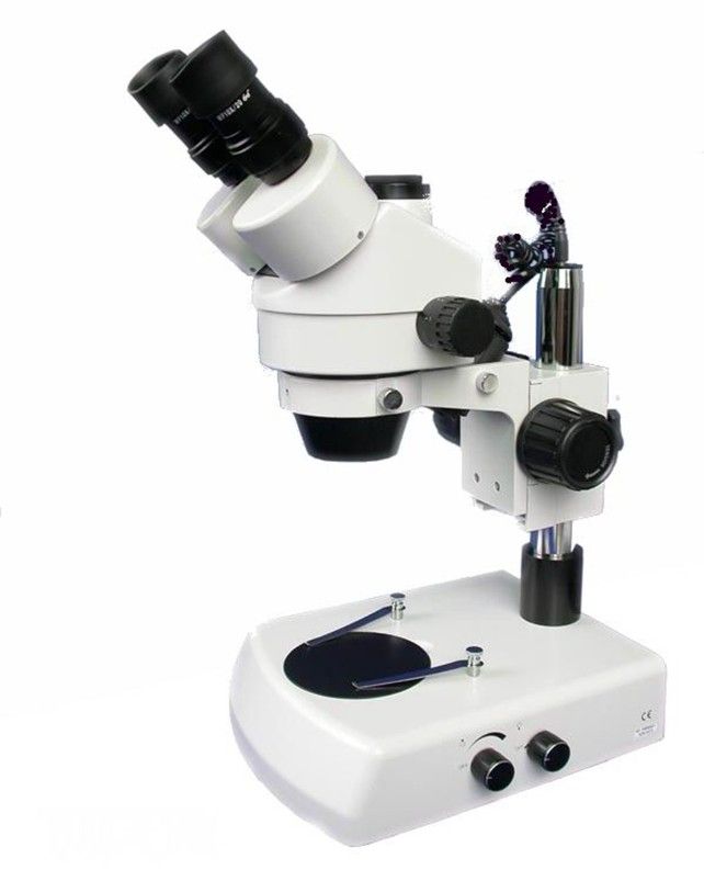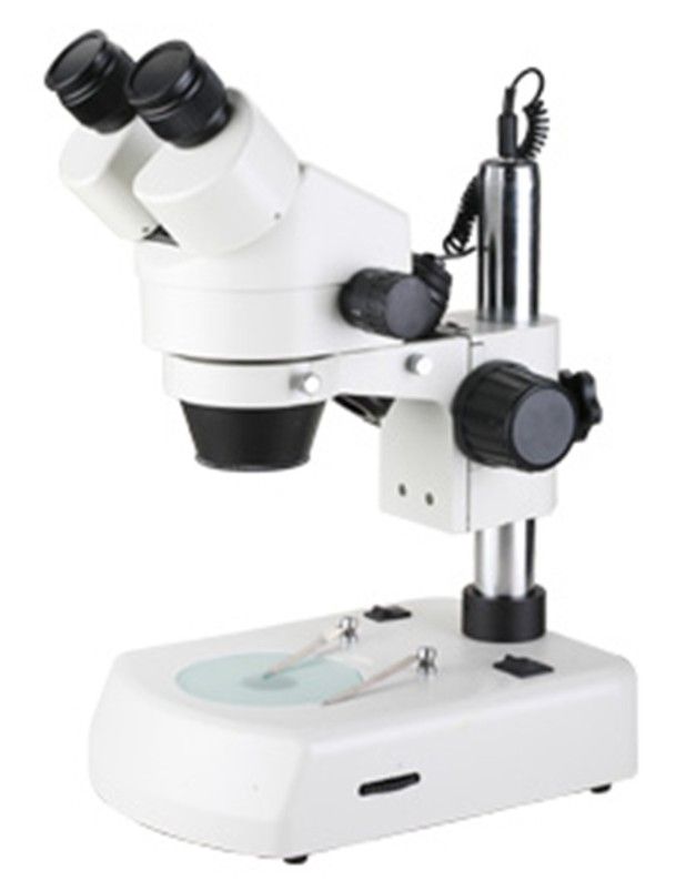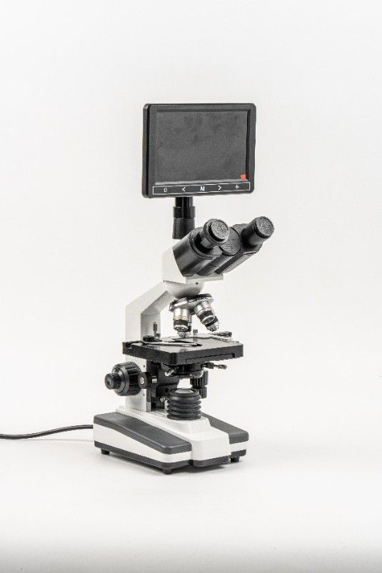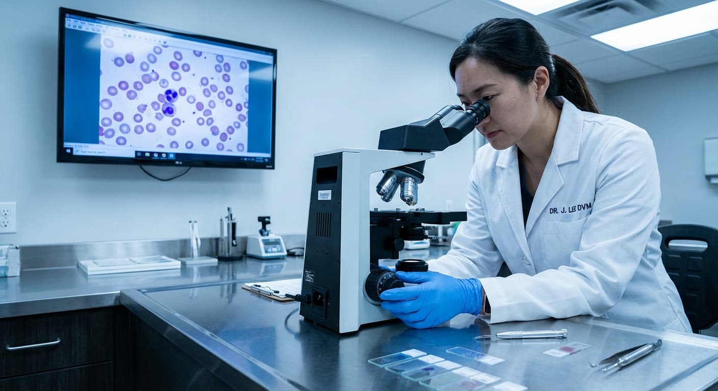
How does a veterinary laboratory work? An in-clinic veterinary lab follows the same steps as a human clinical lab, adapted to different species. Staff collect a sample (blood, urine, faeces, skin scrapings, ear swabs or fine-needle aspirates), prepare it (a centrifuge separates serum or plasma and concentrates urine sediment, and slides are smeared and stained) and analyse it: a hematology analyzer counts blood cells, a biochemistry analyzer measures organ values, a urine analyzer reads test strips, and a biological microscope is used for cytology, parasites, blood smears and urine sediment. Results go into the patient record, and samples that need specialist tests are sent to a reference laboratory. This guide covers the microscope, the instrument behind most of these hands-on checks.
Introduction
In the modern veterinary landscape, the shift from outsourced pathology to in-house diagnostics is not merely a trend; it is an operational imperative for maintaining competitive turnaround times and diagnostic accuracy. At the heart of this transition lies the Biological Microscope. For a veterinary clinic, this instrument is not just a magnifying tool; it is the gatekeeper for dermatology, parasitology, hematology, and urinalysis workflows. However, the market is flooded with optical devices ranging from student-grade units to high-end research platforms. For clinic owners and procurement managers, navigating technical specifications—such as numerical aperture (NA), plan achromatic corrections, and Köhler illumination—can be daunting. Selecting the wrong equipment can lead to increased eye strain for staff, diagnostic ambiguity, and a higher Total Cost of Ownership (TCO) due to premature mechanical failure. This guide provides a comprehensive, engineering-driven analysis of biological microscopes tailored specifically for the Animal Diagnostic Laboratory. We will dismantle the marketing fluff to focus on the optical physics, mechanical stability, and ROI factors that define a professional-grade instrument.
Technical Deep Dive: Engineering Excellence in Veterinary Optics
When evaluating a biological microscope for clinical use, "clarity" is a subjective term. To ensure diagnostic confidence, we must quantify performance through specific optical and mechanical parameters.
The Optical System: Infinity vs. Finite
For a high-throughput veterinary practice, an Infinity Corrected Optical System (ICS) is the superior choice over older finite systems. In an ICS, the light beam is parallel between the objective and the tube lens. This design allows for the insertion of auxiliary components—such as polarizers or epi-fluorescence modules—without altering the magnification or introducing image aberration. This future-proofs your investment, allowing the microscope to evolve with your clinic’s diagnostic capabilities.
Objective Lenses: The Critical Component
The objectives are responsible for the primary image formation. Avoid standard "Achromatic" lenses, which only correct for two colors (red and blue) and suffer from field curvature (blurriness at the edges). * Recommendation: Plan Achromatic or Plan Semi-Achromats. "Plan" indicates a flat-field correction, ensuring the image is in focus from the center to the periphery—critical for scanning cytology slides where marginal details matter. * Numerical Aperture (NA): Look for a 100x oil objective with an NA of at least 1.25. This ensures sufficient resolution to distinguish intracellular bacteria or subtle hemoprotozoa.
Illumination Mechanics: Köhler is Non-Negotiable
Proper illumination is as vital as the glass itself. Köhler Illumination: This system provides even brightness and contrast control. It allows the user to adjust the field diaphragm to match the objective’s aperture, preventing glare and maximizing resolution. Light Source: Transition away from Halogen. 3W LED illumination (5500K–6000K daylight equivalent) is the industry standard. It eliminates the heat transfer associated with halogen bulbs—which can dry out fluid samples (like urine sediments) or kill motility in sperm analysis—and lasts far longer than a halogen bulb.

Comparison Table: Entry-Level vs. Professional Grade
The following table contrasts "Level 1" units often found on general marketplaces against professional-grade units.
Feature | Generic / Level 1 Units | Professional-Grade Units |
|---|---|---|
Optical System | Finite (160mm tube length). Difficult to upgrade. | Infinity Corrected System (ICS). Modular and scalable. |
Objectives | Standard Achromatic. Blurred edges (field curvature). | Plan Achromatic. Flat-field correction for edge-to-edge clarity. |
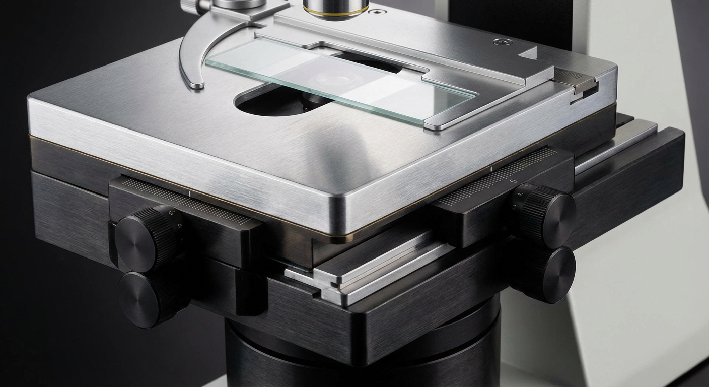
Stage Mechanics | Plastic gears or rack-and-pinion exposed to dust/fluids. Prone to drift. | Rackless / Double-Layer Mechanical Stage. Graphite-treated surface, fluid-resistant, precision movement. |
Illumination | Standard Bulb or low-CRI LED. Inconsistent brightness. | Köhler LED Illumination (3W+). Constant Color Temperature (CCT) regardless of intensity. |
Anti-Fungus | None or superficial coating. | Multi-layer Anti-Fungal Treatment on all optical surfaces (essential for humid lab environments). |
Condenser | Fixed Abbe Condenser (NA 1.25). | Centerable & Focusable Abbe Condenser with iris diaphragm for optimal contrast control. |
TCO (5 Years) | High (Bulb replacements, alignment repairs, user fatigue). | Low (LED longevity, durable mechanics, sustained resale value). |
Clinical Workflow Integration
A biological microscope does not operate in a vacuum; it is part of a high-speed ecosystem.
Ergonomics and Staff Efficiency
Veterinary technicians often spend hours at the microscope. Cumulative trauma disorders (CTD) in the neck and back are real risks. Look for Siedentopf binocular heads with a 30° inclination and 360° rotation. Unlike sliding heads, Siedentopf designs allow interpupillary distance adjustment (usually 48mm–75mm) without changing the tube length or focus. This allows different staff members to switch places instantly without recalibrating the device.
The "Wet" and "Dry" Bench Layout
Positioning is key. The microscope should be situated in the "Dry" area of the lab, away from vibrations caused by centrifuges or the humidity of the Sterilizer (Autoclave). However, it must be proximate to the staining station. A common workflow inefficiency is placing the microscope too far from the Medical Imaging workstations where digital records are stored. Modern microscopes with trinocular heads allow for the mounting of C-mount cameras, enabling live transmission of cytology findings directly to the patient's digital file or a consultation screen.

The Business Case: ROI on Quality Diagnostics
Investing in a premium biological microscope is a financial decision with measurable returns. 1. Revenue Capture: By upgrading optics, a clinic can confidently perform in-house cytology and urinalysis that might otherwise be sent to a reference lab. Offering that service in-house with immediate results improves client compliance and retention. 2. Reduced Diagnostic Error: Poor optics lead to "non-diagnostic" samples or missed artifacts. This necessitates re-sampling (costing staff time and supplies) or, worse, misdiagnosis. High-resolution Plan objectives minimize this risk. 3. Client Education: A trinocular microscope with a camera allows vets to show the client the ear mites or bacteria on a screen. This visual evidence significantly increases treatment plan acceptance rates.
Buying Guide: Procurement Checklist
Procurement managers should demand answers to these three questions before finalizing a purchase order: 1. "Is the stage rackless?" Why ask: Traditional stages have a rack that protrudes from the side when moving the X-axis, creating a safety hazard and a snag point for wires or lab coats. Rackless stages are safer and more durable. 2. "What is the field of view (FN) number on the eyepieces?" Why ask: Standard units offer FN18 (18mm). Professional units offer FN20 or FN22. A wider field of view lets technicians scan slides faster. 3. "Is the system modular for Phase Contrast or Darkfield?" Why ask: Even if you don't need it today, having the slot available for a Phase Contrast slider (ideal for urine sediments and unstained samples) prevents you from having to buy a whole new microscope later.
The HQS Medical Advantage
At HQS Medical, we bridge the gap between biomedical engineering and veterinary application. We understand that a microscope in a vet clinic faces challenges different from one in a university research center—animal hair, dust, and frequent user rotation. For clinic labs, HQS supplies biological microscopes such as the XSZ-107BN, with a compensation-free binocular head inclined at 30°, 4×, 10×, 40× and 100× (oil) objectives, WF10× eyepieces, a double-layer mechanical stage (140 × 140 mm) and coaxial coarse and fine focus. Trinocular models such as the XSZ-107SM add a port for a camera. We do not just sell a device; we provide a cornerstone for your diagnostic capability.
Maintenance & Longevity
To ensure the TCO remains low, strict maintenance protocols are required: * Oil Immersion Hygiene: The 100x objective requires immersion oil. Failure to wipe this lens (and accidentally dipping the 40x lens in oil) is the #1 cause of image degradation. Use only lens paper and manufacturer-approved optical cleaner. * Fungus Prevention: Even with anti-fungal coatings, microscopes should be covered when not in use. In high-humidity climates, storing optics in a dehumidified cabinet is recommended. * LED Management: While LEDs last years, the dimmer pot (potentiometer) is a wear item. Ensure your staff turns the light off when not in use to preserve the electronics.
FAQs
Q: Do I really need a trinocular head if I don't plan to take photos immediately?
A: Yes, we highly recommend it for future-proofing. The cost difference is marginal compared to the cost of replacing the entire head later. As Animal Monitoring & Life Support systems become more digital, integrating microscopy into your Practice Management Software (PMS) will soon be the standard.
Q: What is the difference between Achromatic and Plan Achromatic objectives?
A: Achromatic lenses focus the centre of the field sharply, but the edges may be blurry. Plan Achromatic lenses keep the image in focus across almost the whole field of view. For veterinary cytology where you are scanning the entire slide for abnormalities, Plan Achromatic is essential.
Q: Can I use standard alcohol to clean the lenses?
A: No. High-concentration alcohol can dissolve the cement holding the lens elements together. Use a proprietary optical cleaning solution or a specific mixture of ether and alcohol as recommended in the microscope's user manual.
Q: How often should the microscope be professionally serviced?
A: In a busy veterinary practice, we recommend an annual professional preventative maintenance (PM) visit to lubricate gears, align the optical path, and deep clean the internal prisms.
Conclusion
The biological microscope is a precision instrument that directly influences the speed and accuracy of your veterinary diagnoses. By prioritizing Infinity Corrected Systems, Plan Achromatic optics, and durable LED illumination, you are investing in a tool that enhances clinical outcomes and drives revenue. Avoid the pitfalls of generic "level 1" equipment. Choose a partner that understands the rigorous demands of veterinary medicine. Contact HQS Medical today to discuss configuration options that align with your clinic's specific diagnostic workflow.
Explore Our Biological Microscope Solutions
Interested in learning more about our professional range? Check out our full selection of specialized equipment designed for modern clinics.