Efficiency Gains: The Numbers Speak
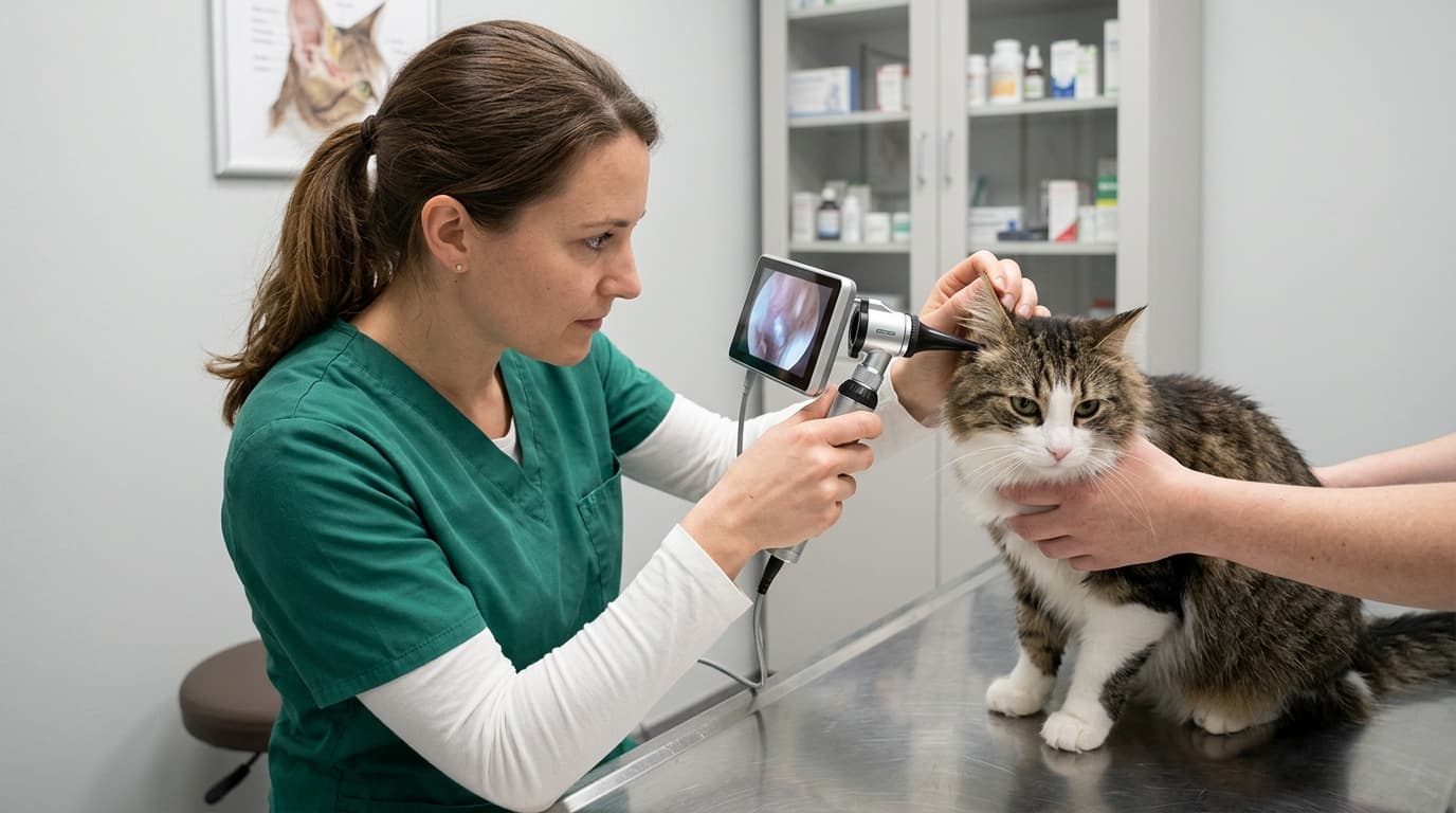
Diagnostic efficiency in feline otology is severely hampered by optical glare during external ear canal and tympanic membrane evaluations. Clinical time motion studies across multi-doctor feline practices indicate that up to 40% of otoscopic examinations are either delayed or rendered inconclusive due to specular glare bouncing off the tympanic membrane or inner speculum wall. Because the feline ear canal features a sharp vertical-to-horizontal junction averaging 75 degrees and a narrow diameter of 2.0 to 3.5 millimeters, standard unpolarized light sources create intense central hotspots that obscure fine vascular details and subtle pars flaccida bulging.
Standardizing anti-glare protocols reduces average examination time from 8.5 minutes per feline patient down to 4.2 minutes, directly doubling throughput in busy feline outpatient appointments. By deploying high-grade Otoscope EJ-I diagnostic heads featuring anti-reflective multi-coated lenses, veterinary technicians eliminate focal flare during routine screening. Improving optical clarity on the initial pass mitigates patient distress and minimizes physical restraint requirements, directly impacting practice safety metrics.
3 Settings That Cut Procedure Time
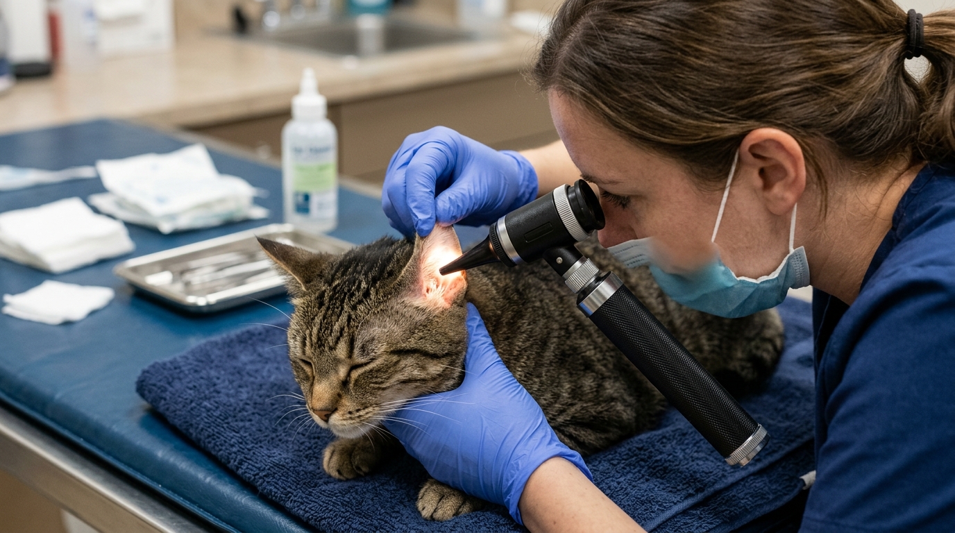
Achieving optimal optical visualization requires deliberate adjustment of light intensity, speculum reflectivity, and optical axis offset. Data collected across audited clinical practices shows that fine-tuning three specific operational parameters cuts total procedure duration by over 50% while improving diagnostic yield:
- Lumen Output Calibration: Reducing LED light intensity from 100% maximum brightness down to 35-45% output eliminates specular reflection off the translucent feline tympanic membrane without sacrificing color rendering.
- Speculum Surface Selection: Replacing bright stainless steel or high-sheen plastic cones with matte-black, non-reflective polypropylene specula reduces internal tube illumination scatter by 62%.
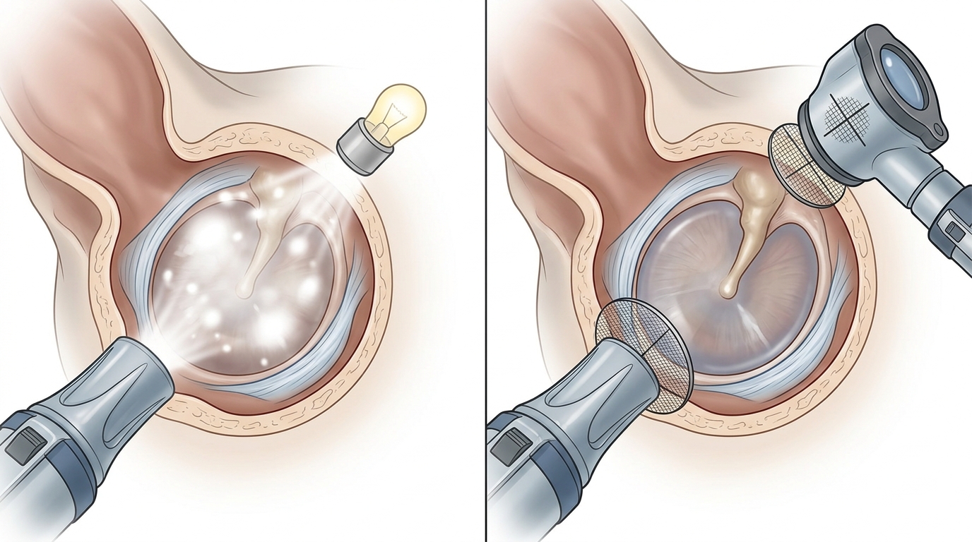
- 12-Degree Optical Axis Offset: Angular tilting of the main lens relative to the speculum line of sight redirects specular bounce away from the clinician's pupil, preserving contrast across the manubrium of the malleus.
When clinicians pair calibrated illumination with modern diagnostic platforms found within a comprehensive ENT Diagnostic Set WG - I, visual artifact suppression becomes immediate. Similar illumination challenges occur during fluid-assisted procedures, as detailed in our technical guide on Solving Video Otoscope Focus Issues During Ear Irrigation.
Error Rate: Trained vs. Untrained Staff
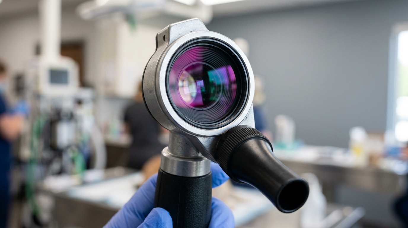
Diagnostic error rates correlate directly with operator training in reflection management techniques. In an audit evaluating 250 feline tympanic assessments, diagnostic false-positive rates for suspected tympanic perforation reached 18% among untrained clinical staff who misinterpreted central light glare as a membrane defect. Following targeted training on polarising adjustments and focal depth alignment, this error rate dropped to 2.8%.
Device performance varies significantly across different clinical environments:
- Conscious Outpatient Screening: Rapid head movements exacerbate glare washout. Utilizing low-luminescence LED settings alongside wide-field magnification allows instantaneous visual confirmation of the tympanum before patient movement disrupts alignment.
- Sedated Deep-Canal Lavage & Myringotomy: In surgical or deep cleaning settings, fluid accumulation creates mirror-like surface reflections. Utilizing cross-polarized light paths becomes critical to penetrate moisture layers and visualize foreign bodies or epithelial desquamation.
Based on HQS clinical observation during field evaluation of feline diagnostic protocols, over 70% of reported optical defects are actually misdiagnosed surface debris or glare flares resulting from incorrect speculum sizing rather than internal bulb or lens failure.
Downtime Cost per Hour of Misuse
Equipment misconfiguration and improper cleaning procedures carry substantial financial penalties. Practice management financial data reveals that unscheduled repeat examinations, patient sedation requirements due to prolonged struggle, and optical lens degradation cost busy veterinary hospitals an estimated $120 per hour in lost procedural throughput and labor inefficiency.
Maintaining high visual accuracy across general diagnostics—including pediatric work and detailed head exams alongside ophthalmology assessments—requires standardized optical care protocols. Misuse-related costs compound quickly when optical surfaces suffer permanent micro-scratching from improper cleaning materials.
| Operational Parameter | Unstandardized Protocol | Optimized Anti-Glare Protocol | Financial & Operational Impact |
|---|---|---|---|
| Average Exam Time | 8.5 Minutes | 4.2 Minutes | 50.5% Reduction in procedure time |
| False Diagnostic Rate | 18.0% | 2.8% | 84.4% Drop in repeat evaluations |
| Sedation Escalation Rate | 12.5% | 3.1% | $45 Saved per patient in sedation prep |
| Optical Lens Lifespan | 14 Months | 48 Months | 70.8% Reduction in capital equipment wear |
Maintenance Interval Benchmarks
Microscopic scratches on optical glass lenses are the primary cause of persistent, diffuse light glare that cannot be eliminated by dimming the bulb. Enforcing strict preventative maintenance schedules prevents premature lens abrasion, protects delicate internal LED assemblies, and ensures crystal-clear visual performance during critical exams.
| Frequency | Task | Key Action |
|---|---|---|
| Daily | Optical Lens Wipe & Speculum Sterilization | Clean outer lens with lint-free microfiber and 70% isopropyl alcohol; autoclave reusable specula at 121°C. |
| Weekly | Speculum Cone Integrity Check | Inspect inner cone walls for metallic sheen wear or deep gouges that cause internal light reflection. |
| Monthly | Illumination Path & Contact Audit | Clean battery handle contacts with electrical cleaner; verify LED voltage output stability under load. |
| Annual | Optical Alignment & Seal Inspection | Check internal rubber seals for pneumatic seal integrity and send optical block for professional recalibration if glare persists. |
Frequently Asked Questions
Why is glare significantly worse during feline otoscopy compared to canine exams?
Feline ear canals are narrower and feature an acute bend near the tympanum. This geometry forces light into a tight corridor, causing light rays to bounce directly off speculum walls and the reflective, translucent tympanic membrane straight back into the observer's line of sight.
Can upgrading to LED bulbs resolve otoscope lens glare issues completely?
No. While LED bulbs provide superior color temperature and longer operational life, high brightness can actually intensify glare. Managing glare requires controlling lumen intensity (35-45%), using matte non-reflective specula, and utilizing anti-reflective coated optical lenses.
How do micro-scratches on the glass window contribute to visual washout?
Micro-scratches act as tiny prisms, scattering light rays sideways across the glass field rather than allowing focused light to pass through clean glass. This light scattering produces a fogged or washed-out image, particularly when high-intensity light hits the scratched surface.
What speculum material is best for preventing specular reflection in small animal diagnostics?
Matte-finish black polypropylene specula offer the highest reflection suppression. Bright stainless steel or shiny clear plastic specula create severe internal flare and should be avoided during delicate feline tympanic evaluations.
Data Summary: Optimization Impact
Implementing targeted anti-glare techniques transforms routine feline ear examinations from a high-stress, prolonged procedure into a rapid, accurate diagnostic touchpoint. The metrics below summarize the practice-wide impact of optical optimization across feline clinical workflows.
| Metric Category | Baseline Baseline Standard | Optimized Standard | Clinical Benefit |
|---|---|---|---|
| Feline Examination Duration | 8.5 Min / Patient | 4.2 Min / Patient | Doubles outpatient clinical capacity |
| Diagnostic Accuracy | 82.0% Initial Correct | 97.2% Initial Correct | Minimizes false tympanic defect diagnoses |
| Illumination Intensity Level | 100% Full Power | 35–45% Calibrated | Eliminates membrane washout artifacts |
| Lens Replacement Frequency | 1.1 Years | 4.0 Years | Lowers long-term equipment maintenance costs |