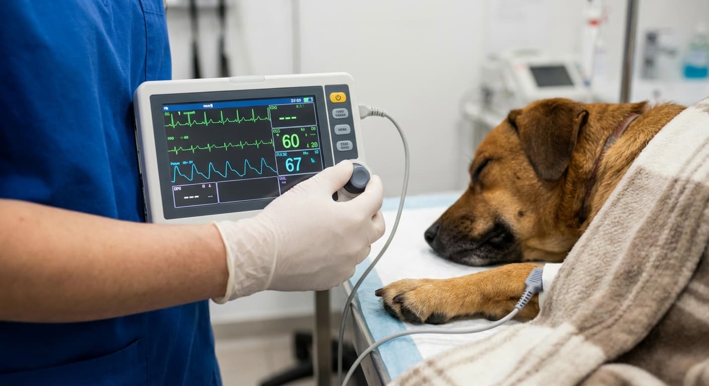
How do you set up a mini veterinary monitor? Charge the battery fully, switch the unit on and select the animal type and patient ID. Choose spot mode for single checks or monitoring mode for continuous recording. Attach the SpO2 clip to thin, unpigmented tissue such as the tongue of an anaesthetised animal, the ear, a toe web, the lip, or the prepuce or vulva. For blood pressure, pick a cuff whose width is about 40% of the limb or tail circumference and place it on a limb or the base of the tail at about heart level. Add the temperature probe, set alarm limits for the species and size of the patient, and wait for a stable pleth waveform before trusting the numbers. On the HQS HM-3T, which measures SpO2, non-invasive blood pressure (NIBP) and temperature, select big- or small-animal mode before measuring so the NIBP range matches the patient.
Maximizing Clinical Outcomes with Precision
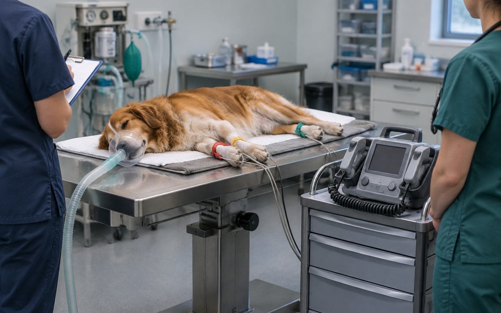
Operational efficiency in veterinary medicine is no longer solely about surgical skill; it is increasingly defined by the integration of diagnostic telemetry. The deployment of a Mini Veterinary Monitor represents a shift towards decentralized, point-of-care diagnostics, allowing practitioners to maintain ICU-level vigilance in anesthesia induction rooms, recovery cages, and mobile field settings. Clinical outcomes correlate directly with the fidelity of physiological data captured during the "golden minutes" of emergency triage and post-operative recovery.
Adopting these compact systems requires a departure from traditional "plug-and-play" mentalities. Many alarms are caused not by the patient but by poorly placed sensors or default settings. Establishing a rigorous setup protocol is essential to filter out artifacts and ensure that the displayed waveform represents the true hemodynamic status of the animal. This guide explores the technical nuance required to elevate a standard Multiparameter Monitor from a passive display screen to an active clinical decision-support tool.
Signal quality depends on how the device filters noise. The HQS HM-3T mini veterinary monitor, for example, uses motion compensation for SpO2 and reports perfusion index (0.05–20%), and its NIBP module uses self-adaptive inflation and deflation algorithms. Getting the most from these features still depends on correct sensor placement and a standard setup routine.
Core Principles of Calibration & Setup
Calibration is the bedrock of diagnostic accuracy. Unlike larger, stationary hospital units, portable monitors are frequently moved, subjecting internal piezoelectric sensors and pneumatic modules to mechanical stress. NIBP (non-invasive blood pressure) modules can drift over time, so their accuracy should be checked periodically. A strict initialization sequence must be followed before every surgical shift.
Zeroing the pressure transducer is non-negotiable for invasive blood pressure (IBP) monitoring, yet often overlooked in EtCO2 (End-tidal CO2) modules. Sidestream CO2 sensors require a clear air calibration to establish a baseline of 0 mmHg partial pressure. Failure to perform this simple step in an environment saturated with exhaled anesthetic gases can lead to "baseline drift," where the monitor reports rebreathing of CO2 that is not physiologically present. This inevitably leads to unnecessary circuit changes and wasted anesthetic gas.
Configuration of alarm limits must be patient-specific rather than device-default. Setting the heart rate low-limit alarm based on the species' specific bradycardia threshold (e.g., 50 bpm for a large dog vs. 140 bpm for a ferret) prevents alarm fatigue. Alarm fatigue causes desensitization, a phenomenon where staff subconsciously ignore audible warnings, posing a severe risk to patient safety.
Clinical Best Practices for Modern Veterinarians
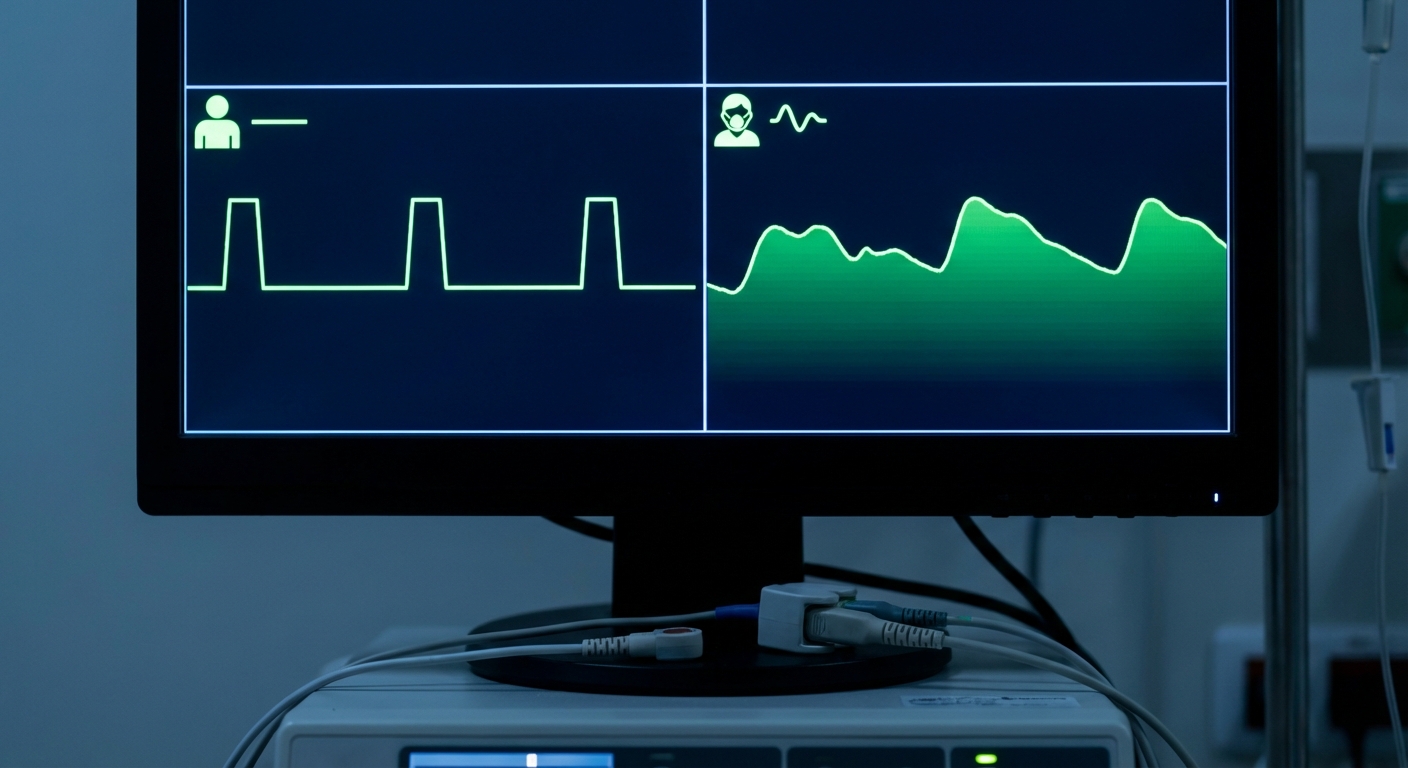
Deploying sensors on veterinary patients involves overcoming anatomical challenges rarely seen in human medicine. Fur density, skin pigmentation, and tissue perfusion variance all affect signal transmission. For SpO2 monitoring, the transmission spectrophotometry principle dictates that the emitter and detector must be perfectly aligned. In animals with pigmented mucous membranes (like Chow Chows or certain Gir cattle), standard red/infrared ratios may yield lower saturation readings. The solution lies in utilizing reflectance probes or placing transmission clips on unpigmented areas such as the vulva or prepuce.
Electrocardiogram (ECG) setup demands rigorous skin preparation. Dry skin and fur act as dielectrics, impeding electrical conduction. The application of 70% isopropyl alcohol to the electrode site decreases skin impedance, but excessive use can cool the patient and cause shivering artifacts. A conductive gel or paste specifically formulated for veterinary use provides a stable electrolyte interface, significantly reducing baseline wander compared to alcohol alone.
Streamlining Clinic Workflow Integration
Data isolation creates bottlenecks. A mini monitor should not function as a data silo. Integration with Practice Management Software (PMS) is the standard for 2026. While many mini monitors operate offline, advanced models support HL7 or DICOM output protocols. Connecting the monitor to the clinic’s local network allows vital signs to be automatically populated into the patient’s digital anesthesia record. This removes transcription errors from manually written anaesthesia charts.
Hardware integration is equally vital. Compact monitors benefit from mounting solutions that position the screen at eye level while keeping cables clear of the surgical field. Utilizing articulating arms or IV pole mounts clears workspace on the operating table, reducing the risk of accidental device drops—the leading cause of premature sensor failure.
Proactive Maintenance & Rapid Troubleshooting
Heat shortens the life of batteries and electronic components. Internal batteries and capacitors are particularly susceptible to heat degradation. Ensuring that the monitor’s cooling vents are never obstructed by drapes or blankets is a critical, zero-cost maintenance strategy.
Cable management requires discipline. Fiber optic cables within SpO2 probes are brittle. Coiling them tightly ("wrapping") fractures the internal glass fibers, leading to intermittent signal loss. The "over-under" coiling technique used in audio engineering should be adopted for medical cables to maintain their internal geometry. Regular cleaning of NIBP Velcro cuffs is also mandatory; accumulated fur and debris reduce grip strength, causing the cuff to pop open during inflation, resulting in "Air Leak" errors and failed readings.
Advanced Technical Mastery for Practitioners
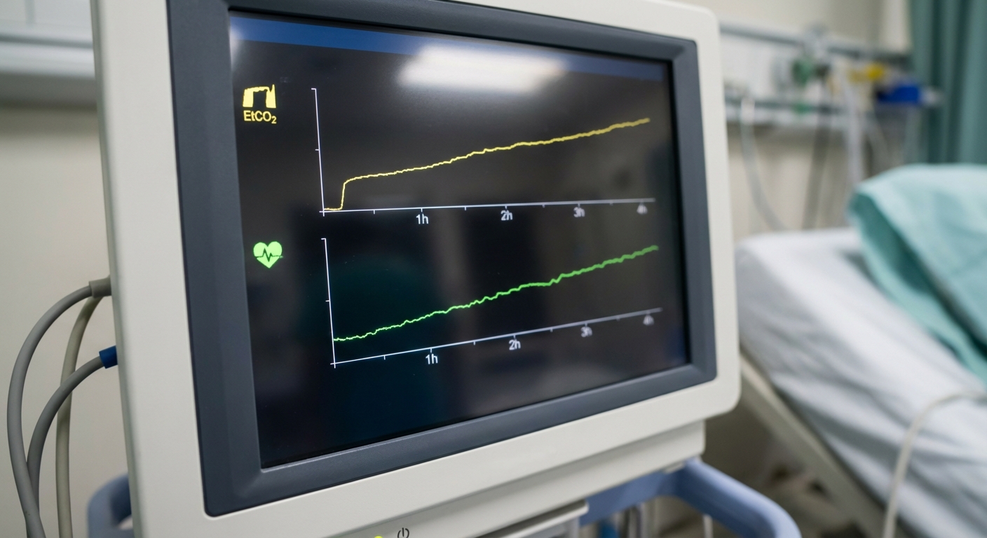
Unlock the hidden potential of the device by utilizing trend data analysis. Most operators focus on real-time numbers, but the 24-hour trend graph is a diagnostic goldmine. It reveals slow-onset trends such as gradual hypercapnia (suggesting exhausted soda lime) or creeping hypotension (suggesting occult hemorrhage) that are invisible in moment-to-moment observation. Configuring the device to display "Mini-Trends" alongside real-time waveforms provides a contextual view of patient stability.
Perfusion Index (PI) is an underutilized parameter available on most modern chipsets. It reflects the pulse strength at the sensor site. A dropping PI is often the first indicator of peripheral vasoconstriction due to shock, hypothermia, or pain, occurring minutes before blood pressure drops. Tracking PI allows the anesthetist to lighten anesthesia depth or administer a fluid bolus pre-emptively.
Frequently Asked Questions
Q: How often should the NIBP module be calibrated on a mini veterinary monitor?
Industry standards dictate that NIBP modules should be verified for pressure accuracy at least once every 12 months, or sooner if the device is dropped. A static pressure test against a calibrated mercury or digital manometer is required to ensure readings remain within ±3 mmHg.
Q: Can mini monitors be used effectively on exotic species like reptiles or birds?
Yes, but specific accessories are required. Standard algorithms are often optimized for cats and dogs. For avians and reptiles, specialized high-sensitivity SpO2 sensors and esophageal ECG probes are necessary to detect the rapid heart rates (often >300 bpm) and low-amplitude signals typical of these species.
Q: Why does the SpO2 reading sometimes fail under bright surgical lights?
Optical interference occurs when ambient light matches the wavelength of the sensor's photodetector. Surgical LED lights can flood the detector, causing signal washout. Covering the sensor site with a small opaque drape or using a purpose-built light shield resolves this issue immediately.
Q: What is the best method to clean the monitor screen without damaging the anti-glare coating?
Avoid ammonia-based cleaners or paper towels, which cause micro-abrasions. Use a microfiber cloth slightly dampened with 70% isopropyl alcohol or a dedicated medical screen wipe. Never spray liquid directly onto the unit to prevent ingress into the bezel seals.
Elevating Standards of Care through Expertise
HQS Medical recognizes that the monitor is only as effective as the operator interpreting it. The Mini Veterinary Monitor is a marvel of engineering density, packing critical diagnostic power into a handheld form factor. By adhering to rigorous setup protocols, understanding the physics of signal acquisition, and maintaining the hardware proactively, veterinary professionals transform this device from a simple gadget into a lifeline.
Mastery of these systems reflects a commitment to the highest standard of animal welfare. As technology advances towards 2026 and beyond, the synergy between skilled clinical judgment and precision instrumentation will continue to define the frontier of veterinary excellence.