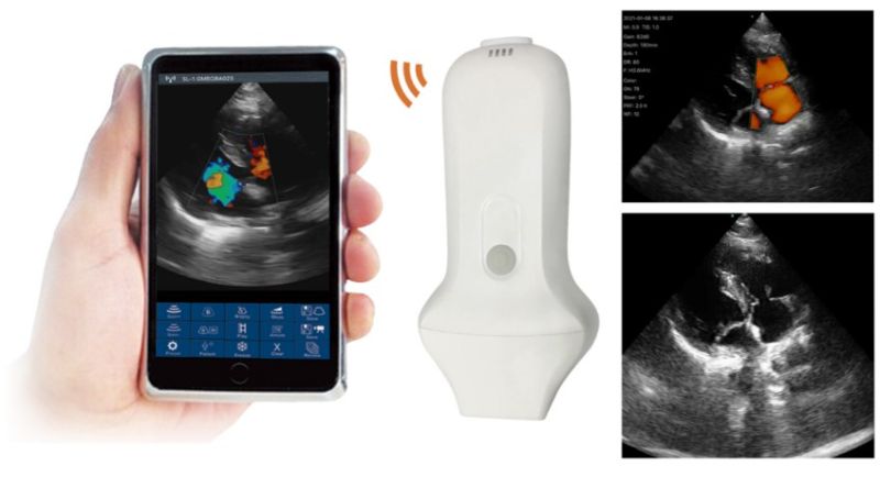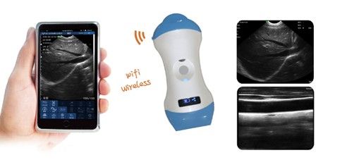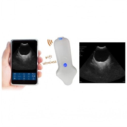
A vet ultrasound purchase goes well when it starts from the examinations you perform, not from a screen size or a brochure headline. This guide explains how to choose a veterinary ultrasound machine for small animal or mixed practice: examinations and species, probes, imaging modes, and portable, cart-based or wireless formats. It ends with a quotation checklist.
You can browse the full range on the HQS ultrasound scanner category page.
Start with the examinations and species
List the examinations you expect to perform most often. Most veterinarian ultrasound work falls into the groups below, and each points to a probe, a depth range and an imaging mode.
- Small animal abdomen: liver, spleen, kidneys, bladder, intestines and lymph nodes in dogs and cats. This is the core of pet ultrasound diagnostics and usually calls for a convex or micro-convex probe.
- Cardiac examinations: chamber size, wall motion and valve flow. Echocardiography needs a small footprint to image between the ribs, M-mode for timing and measurements, and spectral Doppler for velocities.
- Reproduction: pregnancy checks in dogs and cats, and rectal scanning of cattle and horses. Farm work favors low weight, battery operation and a rectal probe over a large display.
- Superficial structures: tendons, eyes, thyroid, skin masses and guided sampling, where a higher-frequency linear probe gives finer detail.
A mixed practice may need two tools: a console for the clinic and a portable unit for farm calls.
Veterinary ultrasound probe types in general terms
The probe decides image quality as much as the main unit does. Higher frequencies give finer resolution but less penetration; lower frequencies reach deeper with less detail. Common probe shapes:
- Convex (curvilinear): a wide, fan-shaped field of view for the abdomen.
- Micro-convex: the same fan shape on a smaller footprint, useful for cats, small dogs, exotic species and scanning between ribs.
- Linear: a rectangular field at higher frequency for superficial structures, tendons, small parts and vascular access.
- Phased array: a very small footprint that steers the beam electronically, the usual choice for echocardiography through intercostal windows.
- Rectal (transrectal): a long, slim probe designed for reproductive scanning in cattle and horses.
Treat each veterinary ultrasound probe as a separate line item. Ask which probes are included, which are optional, and whether each supports every listed mode.
Imaging modes: B, M, color Doppler and spectral Doppler
B-mode and M-mode
B-mode is the standard grayscale image and covers most abdominal and reproductive work. B+B shows two images side by side for comparison. M-mode plots tissue movement along one line over time, for cardiac measurements and fetal heartbeat.
Color and power Doppler
Color flow mapping (CFM) overlays the direction and mean velocity of blood flow on the B-mode image. Power Doppler (PDI) displays the strength of the flow signal rather than its direction, which makes it more sensitive to slow flow in small vessels. Directional power Doppler (DPDI) adds direction information to the power display. For color Doppler veterinary work in cardiology and vascular assessment, these modes are the reason to move up from a B/W system.
Spectral Doppler: PW, HPRF PW and CW
Pulsed-wave (PW) Doppler measures velocity at a chosen depth, up to a limit above which the signal aliases. High pulse repetition frequency PW (HPRF PW) raises that limit. Continuous-wave (CW) Doppler measures high velocities along the whole beam line without depth selection, which is why it matters for fast cardiac jets. If echocardiography is on your list, confirm that CW is enabled on the probe you will use for cardiac work.
Why pseudo-color is not Doppler
Many B/W scanners offer a pseudo-color function that maps grayscale values to a color palette. It carries no flow information, so a colored brochure image proves nothing about Doppler. Check the mode list for CFM, PDI, PW or CW by name.
Portable, cart-based or wireless vet ultrasound?
Format is the second decision after the examination list.
- Handheld units with their own screen are light and suit farm rectal scanning and quick clinic checks. Screens are smaller and mode lists shorter.
- Cart or console systems have larger displays, more probe connections and fuller Doppler packages.
- Wireless probes send images to a separate display device. They remove the cable but depend on a compatible device, the app and battery life. Confirm which phone or tablet is supported and whether it is supplied.
A portable veterinary ultrasound unit is rarely a direct substitute for a console in cardiology, but it is often the right tool for reproduction and field work.
Comparing three HQS veterinary ultrasound machine formats
The three models below represent three formats. Shortlist the format first, then specify probes, modes and image storage in your quotation.
| Model | Type | Display | Listed modes | Other listed facts |
|---|---|---|---|---|
| US-S1 | Handheld digital B/W ultrasound scanner | 5.6 inches | B, B+B, B/M | Brochure standard package: 6.5 MHz rectal probe; other probes optional |
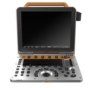
| US-P60 | Color Doppler ultrasound system | 15-inch LCD | Color: CFM, PDI, DPDI. Spectral: PW, HPRF PW, CW | Probes and enabled modes depend on the quoted configuration |
| US-3V | Wireless transrectal probe ultrasound scanner | Separate display device (confirm compatible device and app) | B, B/M | Published weight: 600 g. Image formats: JPG, AVI, DICOM |
The US-S1 suits reproductive scanning when its probe matches your examination list; confirm the included probe, battery, memory card and adapter.
The US-P60 is the format to consider when cardiac and vascular work matter. Ask for the exact probes and enabled modes in writing, with sample images from that configuration.
For the US-3V, confirm the compatible display device and app, the quoted probe configuration and the export workflow.
Other scanners in the range include the US-S10, US-3ML and US-4PA. Ask for their specification sheets if their format fits your case list.
Image storage and export
Images that cannot leave the machine are hard to use for referrals and records. Ask:
- Which still and video formats are supported, for example JPG for stills and AVI for clips.
- Whether DICOM is listed for the exact model, and whether it covers sending to a PACS or only file export.
- How images are transferred: memory card, USB, network or app.
- How patient identification is entered so that files can be matched to records.
Treat DICOM as available only when it is listed for the model you are buying. Among the three models above, it is listed for the US-3V.
Positioning and probe care
A stable surface that is easy to clean makes positioning faster. The ZL-03 ultrasound examination table is a stainless steel table for B-mode examinations with rounded edges and universal brake wheels. It measures L110 × W55 × H70 cm, and the size can be customized.
Probe care habits:
- Wipe off gel after every patient with a soft, non-abrasive cloth.
- Clean and disinfect only with agents the manufacturer lists as compatible.
- Use probe covers for rectal work and follow the manufacturer's disinfection instructions for those probes.
- Inspect the lens, housing and cable for cracks, and stop using a damaged probe.
Request the written probe-cleaning instructions and details of replacement-probe support together with the quotation.
What to put in your quotation request
- Your examination list, species and animal sizes.
- The exact model and format: handheld, console or wireless.
- Every included probe by type and frequency, plus optional probes.
- The enabled imaging modes, naming CFM, PDI, PW, HPRF PW or CW where needed, and any software licenses.
- For wireless devices: the supported phone or tablet, the app, and whether a display device is supplied.
- Battery, memory card, adapter and other accessories in the package.
- Image formats and export workflow, including DICOM where listed.
- Sample images from the configuration being quoted.
- Probe-cleaning instructions and replacement-probe support.
Cost depends mainly on the format, the number and type of probes, and the enabled Doppler modes. A vet ultrasound machine quotation built on this list lets you compare offers on equal terms. Send your requirements through the ultrasound scanner category or the model page that fits your practice.
Frequently asked questions
Which probes does a dog ultrasound machine need?
For the canine abdomen a convex probe is the usual starting point, with a micro-convex probe for small dogs and cats. Add a phased array probe for echocardiography and a linear probe for superficial structures.
Is a portable veterinary ultrasound enough for cardiac scanning?
Echocardiography needs a small-footprint probe, M-mode and spectral Doppler, including CW for high-velocity flow. Many portable B/W units do not list these modes, so check the named modes before buying. Scanning protocols and interpretation remain decisions for the veterinary team.
What should a veterinary sonography machine for cattle and horses include?
A rectal probe, battery operation, low weight and a display you can read in barn conditions. The US-S1 standard package lists a 6.5 MHz rectal probe, and the US-3V is a wireless transrectal probe device with a published weight of 600 g.
Does a colored image mean the scanner has Doppler?
No. Pseudo-color maps grayscale values to colors and shows no blood flow. Doppler capability is shown by named modes such as CFM, PDI, PW or CW in the specification of the exact model and configuration.
Can animal ultrasound images be stored in a PACS?
Only if DICOM is supported for the model and configuration you buy. Ask whether it covers network sending or only file export.