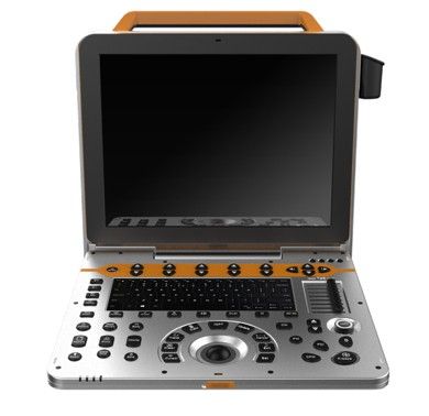
Color Doppler Ultrasound Scanner
Introduction to Color Doppler Ultrasound Scanner
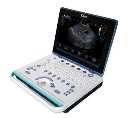
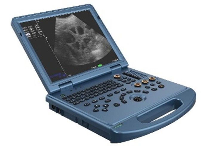
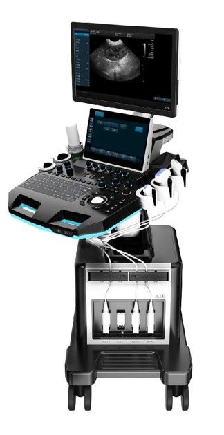
The US - P60 Color Doppler Ultrasound Scanner is designed for the examination and diagnosis of the digestive, reproductive, and urinary systems in animals. It is suitable for use in veterinary hospitals and scientific institutions, featuring a high-resolution LCD monitor and advanced imaging capabilities.
Key Features of US - P60
- Applicable for animals’ examination and diagnosis on digestive system, reproductive system, urinary system for animal hospitals and scientific institutions.
- 15 inch high resolution medical LCD monitor.
- Anatomic M Mode Imaging (Sampling line≥ 3).
- Contrast Tuned Imaging.
- Tissue Harmonic Imaging (THI).
- B/C/D Real - time three synchronous imaging.
- Color Doppler Imaging (CFM, PDI, DPDI).
- Spectral Doppler Imaging (PW, HPRF PW, CW).
- Support: Chinese/ English/ Spanish/ French/ Russian.
- Space compound imaging (application to Abdominal, GYN, vessel, superficial small organs, can be dual image contrast display at same time).
- Imaging including velocity, velocity variance, energy, direction energy display, etc
- Display mode B/C, B/C/M, B/POWER, B/C/PW
- Color hiding technology without back to B mode can hide C mode, only color speed ruler display
- Blood flow graph, color flow profile to measure intravascular velocity
- Display full screen, duplex / triplex (PW only)
- Max scanning speed PWD: Positive or negative blood flow velocity≥7.6m/s ; CWD: blood flow velocity ≥20.0m/s, min ≤5 mm/s (Nonnoise signal)
- Display mode B, PW, B/PW, B/C/PW, B/CW, B/C/CW, etc
- Display control reverse, zero shift, B refresh, D extension, B/D extension, etc
- Smart Doppler technology freely switch between real - time B+CFM mode and real - time PW mode
- Zero shift adjustable ≥8 level
- Automatic Spectrum Measurement, Manual Measurement
- PView W ide F ield I maging
- High - resolution display, length can up to 50cm
- Imaging support forward erasure, no need to re - imaging
- With two dimensional wide view imaging mode and color wide view imaging mode
- Wideband with frequency conversion, independent frequency conversion under B and CFM.
Color Doppler Ultrasound Scanner Specifications
PARAMETER | SPECIFICATION |
|---|---|
2D Imaging Mode | Receiving mode transmitting and receiving channel ≥1024, multi - signal parallel processing |
- | Line density ≥512, visible and adjustable |
- | TGC 8 segment adjustable |
- | Gain B / M / D separately adjustable, ≥100dB |
- | Dynamic range ≥180dB |
- | Max scanning depth ≥360mm |
- | Grey scale 0 - 67, visible and adjustable |
- | Sound power 1% - 100% |
- | Full Digital Beamformers |
Convex probe | Fundamental
Frequency: 2.0MHz/ 2.3MHz/ 2.5MHz/ 3.0MHz/ 3.5MHz/ 4.0MHz/ 4.6MHz/ 5.0MHz/ 5.4MHz (9 levels frequency conversion) |
- | Harmonic
Frequency: 4.0MHz/ 4.6MHz/ 5.0MHz (3 levels frequency conversion ) |
Linear probe | Fundamental
Frequency: 4.0MHz/ 4.6MHz/ 5.0MHz/ 6.0MHz/ 7.0MHz/ 8.0MHz/ 9.2MHz/ 10.0MHz/ 12.0MHz/ 13.3MHz (9 levels frequency conversion) |
- | Harmonic
Frequency: 8.0MHz/ 9.2MHz/ 10.0MHz (3 levels frequency conversion ) |
Micro - convex probe | Fundamental
Frequency: 3.0MHz/ 3.5MHz/ 4.0MHz/ 5.0MHz/ 5.4MHz/ 6.0MHz/ 7.0MHz/ 8.0MHz (8 levels frequency conversion) |
- | Harmonic
Frequency: 6.0MHz/ 7.0MHz/ 8.0MHz (3 levels frequency conversion ) |
Phased array probe | Fundamental
Frequency: 1.7MHz/ 1.9MHz/ 2.1MHz/ 2.5MHz/ 3.0MHz/ 3.4MHz/ 3.8MHz/ 4.2MHz/ 5.0MHz (9 levels frequency conversion) |
- | Harmonic
Frequency: 3.4MHz/ 3.8MHz/ 4.2MHz (3 levels frequency conversion ) |
High frequency phased array probe | Fundamental
Frequency: 3.0MHz/ 3.5MHz/ 4MHz/ 5MHz/ 5.4MHz/ 6MHz/ 7MHz/ 8MHz (9 levels frequency conversion) |
- | Harmonic
Frequency: 6MHz/ 7MHz/ 8MHz (3 levels frequency conversion ) |
Technical requirements for electronic convex probe | Frequency: 2.0 - 5.4 MHz, lateral resolution: ≤2 (depth≤80), ≤3mm (80 < depth≤130) |
- | A xial resolution: ≤1 (depth≤80), ≤1mm (80 < depth≤130) |
- | D ead zone: ≤3mm; Geometric position accuracy: broadwise: ≤5%; lengthwise:≤5% |
- | D etecting depth ≧ 240mm |
Technical requirements for electronic linear probe | Frequency: 4.0 - 13.3MHz, lateral resolution: ≤1 (depth ≤40), |
- | A xial resolution: ≤0.5 (depth ≤50), |
- | D ead zone: ≤3mm; Geometric position accuracy: broadwise: ≤4%; lengthwise: ≤2% |
- | D etecting depth ≧ 100mm |
Customer Reviews(0)
Related Products

Wireless Ultrasound Scanner
Model: US - 3C/ US - 3L

Color Doppler Ultrasound Scanner
Model: US-T5

Color Doppler Ultrasound Scanner
Model: US - L6

Color Doppler Ultrasound Scanner
Model: US - E80
Frequently Asked Questions (FAQ)
What are the key features of the Color Doppler Ultrasound Scanner?
The Color Doppler Ultrasound Scanner features Applicable for animals’ examination and diagnosis on digestive system, reproductive system, urinary system for animal hospitals and scientific institutions., 15 inch high resolution medical LCD monitor., Anatomic M Mode Imaging (Sampling line≥ 3).. It is designed for professional veterinary use and meets international quality standards.
What types of veterinary clinics is the Color Doppler Ultrasound Scanner suitable for?
The Color Doppler Ultrasound Scanner is suitable for small animal clinics, large animal hospitals, emergency veterinary centers, and specialty practices. It can be configured to meet different clinical requirements.
What is the warranty period for this equipment?
All HQS Medical veterinary equipment comes with a standard 12-24 months warranty and lifetime technical support.
Do you offer global shipping?
Yes, we provide fast and secure shipping to over 50 countries worldwide, including all necessary customs documentation.