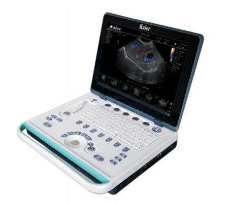
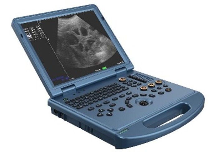
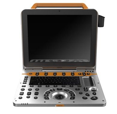
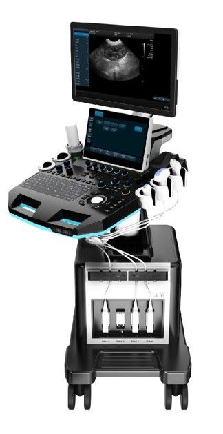
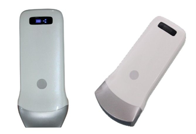
Operating system | Adopts ARM chip architecture, stable, concise and powerful |
Display | 15 - inch 1024*768 resolution high - definition L CD screen |
Display mode | B, B+B, B+M, M, 4B |
Probe array element | The host can automatically identify and use a variety of array element probes (80 array elements.96 array elements.128 array elements) |
Image preset | This machine combines a large amount of clinical experience, has different preset adjustment conditions for different species, and has a one - key optimization function |
Scanning angle adjustment | Adjustable |
Image magnification | Separate knob |
Depth adjustment | ≥20 levels adjustable |
Puncture | With puncture guide line, angle and position are adjustable |
Focus | The focus position is adjustable, the focus position adjustment is ≥5 gears |
Movie playback | ≥256 frames |
Image adjustment | Up and down, left and right, brightness, focus position, dynamic range, scanning angle, frame correlation, M speed |
Image storage | ≥300 frames. External USB storage, single image storage time is about ≤9 seconds (storage speed is related to the USB device used) |
Image processing | Image smoothing/sharpening, tissue harmonics, gamma correction, false color |
Notes and characters | Date, clock, name, age, gender, doctor, hospital name, image annotation |
Position mark | 24 kinds |
Measurement function | a) Routine measurement: distance, perimeter, area, volume; b) Heart measurement: left atrium, right atrium, left ventricle, right ventricle, aorta, ascending aorta, descending aorta, aortic isthmus; c) Obstetric measurement: gestational age (double parietal diameter, head and tail length, gestational sac, head and neck diameter, stomach long axis, heart long axis, skull diameter, trunk diameter, uterine diameter), yolk sac, abdomen, large brain, amniotic fluid, Fibula length, trunk d) Abdominal measurement: liver, gallbladder, kidney, abdominal aorta, iliac artery e) Superficial measurement: thyroid, sperm cyst, testis, breast mass, skin mass And has different species measurement formulas |
Report function | Hospital standard report page settings, you can enter information such as animals, physicians, diagnosis results, etc., and automatically import selected diagnosis pictures and inspection measurement data |
External interface | VGA interface, USB2.0 interface, RS232 interface |
Power saving mode | The panel button light can be turned on and off with one button, and the instrument will automatically turn off the power if it is not used for 6 minutes. Press any key to resume operation |
Probe | 80 element R60 |
Detection depth | ≥160mm |
Lateral resolution | mm ≤3(depth≤80)≤4(8 < depth≤130) |
Axial resolution | mm ≤2(depth≤80)≤3(80 < depth≤130) |
Blind area | mm ≤5 |
Horizontal geometric position accuracy | % ≤15 |
Longitudinal geometric position accuracy | % ≤10 |
Probe slice thickness index | mm ≤10 |
Perimeter and area measurement deviation | % ± 20 |
M mode time and distance error | ± 10 |
Probe working sound frequency deviation | The deviation between the working sound frequency of the probe and the nominal frequency of the machine should be within ± 15% |
Grayscale | 256 levels |
Power supply range | AC 100V ~ 240V, tolerance ± 10%, 50Hz/60Hz, tolerance ± 1Hz. DC 14V ± 0.5V, 3A |
Continuous working time | ≥8 hours |
Battery pack standby time | ≥2 hours |