A veterinary X-ray machine is not a single device. It is a set of parts that have to match each other and the room: a generator and tube that produce the exposure, a detector that captures the image, a table or rack that holds the patient and tube in position, and software that turns raw data into a study the veterinarian can read. This guide explains each part, how to read generator figures, which format suits which caseload, and what to send in an enquiry.
The parts of a veterinary X-ray setup
Generator and tube
The generator converts mains power into the high voltage that drives the X-ray tube. High-frequency designs give a steadier output than older transformers and make short exposures more repeatable. The tube produces the beam. Its focal spot size affects sharpness: a small focal spot gives finer detail on extremities and small patients, while a larger one tolerates the higher loads needed for thick body regions.
Detector
The detector records the X-rays that pass through the patient. In a digital system this is usually a flat panel. Compare three figures: active area, pixel pitch in micrometres, and spatial resolution in line pairs per millimetre (lp/mm). Bit depth describes how many grey levels the panel records. A 16-bit panel leaves room to adjust brightness and contrast after the exposure.
Table, rack or stand
The mechanical part fixes the source-to-image distance (SID) and decides how easily staff can position the animal. Options range from an integrated imaging table to a mobile rack, a C-arm or a small dental arm.
Workstation and software
Acquisition software controls the detector, applies image processing and stores studies. Ask which computer is supplied, whether images export in DICOM format, and whether studies can be sent to a PACS or a referral centre.
DR vs CR vs film
Three capture methods are still found in practices:
- Film needs chemical processing and a darkroom, and every repeat costs time and consumables.
- Computed radiography (CR) uses reusable phosphor plates in cassettes. After the exposure, each plate is scanned in a reader. CR works with an existing generator but adds a plate-handling step to every image.
- Direct radiography (DR) uses a flat-panel detector that sends the image to the computer within seconds. There is no cassette to carry or scan.
The practical gain of veterinary DR X-ray is speed at the table. The image appears while the patient is still positioned, so a repeat view can be taken immediately if positioning was off. The trade-off is that the detector is a valuable component and must be handled with care.
Reading generator output: kW, kV, mA and mAs
Generator specifications use a small set of terms:
- kV (kilovoltage) sets beam energy and penetration. Thick regions such as a large dog's abdomen need more kV than a cat's paw.
- mA (tube current) sets how many X-rays are produced per second.
- Exposure time is given in milliseconds or seconds.
- mAs is mA multiplied by time. It controls the total quantity of radiation and therefore image density and noise.
- kW is the power rating. It limits which combinations of kV and mA the generator can deliver at the same moment.
Power matters because patients move. A panting dog needs a short exposure, and a short exposure at a given mAs requires high mA. A higher kW rating lets a veterinary X-ray generator supply more mA at the kV that large patients need. Compact 5 kW generators are commonly chosen for small animals, limbs and routine studies. Chest and abdomen work on giant breeds pushes toward higher mA or longer times, so check that the listed mA and mAs ranges fit your largest regular patients. Exposure charts and radiation protocols remain decisions for the veterinary team.
Choosing a veterinary X-ray machine format
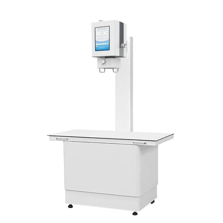
Integrated DR table system
A table system suits a clinic with a dedicated imaging room and a steady small-animal caseload. Compared with separate x-ray tables and stands, an integrated design keeps tube, detector and tabletop aligned. The DR-A10 veterinary digital radiography system is built around a 1210 × 610 mm tabletop with a removable X-ray head and a 17 × 17 in amorphous-silicon detector with 139 µm pixels. Its generator lists 40–120 kVp and 10–100 mA, and the SID is 1000 mm.
Mobile DR rack and portable generator
A mobile system moves to the patient. The DR-100 digital radiography system pairs a compact 5 kW generator on a mobile rack with a 35 × 43 cm CsI detector (140 µm pixels). The generator lists 40–125 kV and 10–100 mA, the generator unit weighs 15 kg, and the set is intended for limb and bedside radiography.
For portable X-ray veterinary work, the RX-100 portable radiography X-ray system is a 5 kW generator weighing 15 kg (365 × 240 × 210 mm) on a 220–240 VAC supply. It covers 40–125 kV in 1 kV steps, 10–100 mA, 1–400 ms and 0.1–200 mAs, with 0.6 / 1.8 focal spots. Options include a mobile rack, electric triangle bracket, T-type trolley, folding trolley or table, and it is listed mainly for limb examinations. Confirm the detector and software you plan to pair with it.
The DR-A10, DR-100 and RX-100 each list three exposure controls: remote, hand switch and touch screen. A remote or hand switch lets the operator stand back from the beam during the exposure.
C-arm fluoroscopy
Fluoroscopy shows a live image during fracture reduction, implant placement or foreign-body retrieval. Pulsed mode images in short bursts and lowers dose compared with continuous mode. For C-arm veterinary use in theatre, the DR-112D mobile C-arm radiography system offers continuous and pulsed fluoroscopy plus radiography from a 3.5 kW high-frequency generator (inverter ≥40 kHz). It uses a 9-inch three-field image intensifier (4.8 lp/mm at the centre), a 1024 × 1024 camera and a 23.8-inch monitor. The C-arm opening is 760 mm with a 640 mm arc depth and a 400 mm electric lift, and the system includes DICOM browsing and network functions.
Dental X-ray
Whole-body radiographs show overlapping structures in the skull, so tooth roots and periodontal bone need intraoral images. A dental radiography machine is a small tube on an arm or in the hand, used with an intraoral sensor or phosphor plates; confirm which capture method is included. The X-10P portable high-frequency dental X-ray lists 60 kV, 2 mA, 0.2–3.2 s exposure, a 14.8 V 6600 mAh battery, an LCD and memory of exposure parameters. The X-10D dental X-ray sits on a mobile or fixed base and lists 70 kVp, 8 mA, 0.2–4 s exposure, a 220 V supply and 50 kg net weight.
HQS X-ray models compared
| Model | Format | Generator | Image capture | Main use |
|---|---|---|---|---|
| DR-A10 | DR table system | 5 kW; 40–120 kVp; 10–100 mA | 17 × 17 in detector, 139 µm pixels | Dedicated imaging room |
| DR-100 | Mobile DR rack | 5 kW; 40–125 kV; 10–100 mA | 35 × 43 cm CsI detector, 140 µm pixels | Limb and bedside radiography |
| RX-100 | Portable generator | 5 kW; 40–125 kV; 10–100 mA; 0.1–200 mAs | Confirm detector in quotation | Mainly limb examinations |
| DR-112D | Mobile C-arm | 3.5 kW high-frequency | 9-inch image intensifier, 1024 × 1024 camera | Fluoroscopy and radiography |
| X-10P | Portable dental | 60 kV; 2 mA; battery | Confirm sensor or plates | Dental radiography |
| X-10D | Dental on base | 70 kVp; 8 mA; 220 V | Confirm sensor or plates | Dental radiography |
The DR-A10 and DR-100 both list a 5 kW generator, 16-bit conversion and 3.6 lp/mm spatial resolution. Full specification tables are on each model page and in the veterinary digital X-ray category.
Room planning and radiation safety
Room shielding, radiation safety, staff monitoring and registration of X-ray equipment follow local regulations, so involve your radiation protection adviser or the competent authority before you order. Measure the room and doorways, decide where the operator will stand, and confirm the mains supply. Plan space for the workstation, a network connection and storage for the detector and aprons.
What drives veterinary X-ray cost
Two quotations for "an X-ray" can describe very different systems. The main cost drivers are:
- Detector size and type, and whether one or two detectors are included.
- Generator power and kV, mA and mAs ranges.
- Mechanical format: integrated table, mobile rack, C-arm or dental unit.
- Acquisition software, workstation, DICOM and PACS connection.
- Installation, commissioning and staff training.
- Room shielding and building work, which are usually arranged locally.
- Accessories such as positioning aids, grids and protective wear, plus shipping.
Ask for a configured quotation that lists every item, so offers can be compared line by line.
What to put in your enquiry
- Species and weight range, and the body regions you image most.
- Your current setup: film, CR or no X-ray yet.
- Room dimensions and mains voltage at the planned position.
- Preferred format: table, mobile rack, portable unit, C-arm or dental.
- Required detector size and software needs, including DICOM export and PACS.
- Accessories wanted and the destination country, so documents for local registration can be prepared.
You can review the full range in the veterinary digital X-ray systems section and send these details with your request.
Frequently asked questions
How powerful does a veterinary X-ray generator need to be?
It depends on your largest regular patients. Small-animal and limb work often uses compact 5 kW units; thick chest and abdomen studies on large dogs need higher mA, so compare kW with the mA and mAs ranges.
Is veterinary digital radiography faster than CR?
Yes. A DR detector sends the image to the computer within seconds, while CR plates must be carried to a reader and scanned.
What affects veterinary X-ray cost the most?
Detector size and type, generator output, mechanical format, software and installation. Shielding and room work are separate local costs.
Can a portable vet X-ray machine replace a table system?
For limbs, bedside work and ambulatory visits, a portable or mobile unit is practical. For many chest, abdomen and spine studies in one room, a table system gives more consistent positioning.
Do I need a separate dental radiography machine?
Yes, if you perform dentistry. Intraoral views need a dental tube with intraoral sensors or plates.