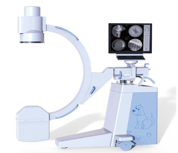
Mobile C-arm Radiography System
Introduction to Mobile C-arm Radiography System
The DR - 112D Mobile C-arm Radiography System offers advanced imaging capabilities with a high-quality X-ray generator and intelligent features for enhanced diagnostic accuracy. Its user-friendly interface, ergonomic design, and robust configuration make it an efficient solution for various orthopedic and surgical applications.
Key Features of DR - 112D
- High quality combined high - frequency and high - voltage X - ray generator, greatly reducing the X - ray exposure.
- Automatic tracking of perspective kV and Ma for optimal image brightness and clarity.
- Human graphical LCD touch screen interface for intelligent and convenient operation.
- Hand-held controller for convenient instrument operation.
- 9-inch three field image intensifier with stable quality and good image definition.
- Megapixel ultra-low illumination digital camera for clearer images.
- Standard workstation and advanced image software processing technology for clearer images and convenient diagnosis.
- Standard DICOM interface for easy integration with hospital information systems.
- New frame design with a small and beautiful appearance.
- Digital photographing function for convenient operation and efficient digital image processing.
- Appliant to Orthopaedics (Osteopathy, Diaplasis, Nailin) & Surgery (orthopedic, removing foreign body, implanting pace maker, partial radiography, local photography, and other work).
Mobile C-arm Radiography System Specifications
Specification | Value |
|---|---|
Monoblock Focus | 0.3/1.5 |
Anode capacity | 35kJ ( 47kHu ) |
Tube Heat capacity | 650kJ ( 867kHu ) |
Power output | 3.5kW |
Inverter Frequency | ≥ 40kHz |
Continous Fluoroscopy (Manual/automatic) Tube voltage | 40kV ~ 110kV |
Continous Fluoroscopy (Manual/automatic) Tube current | 0.3mA ~ 4mA |
Continous Fluoroscopy (Manual/automatic) Automatic brightness tracking function | Yes |
Pulse fluoroscopy Tube voltage | 40kV ~ 110kV |
Pulse fluoroscopy Tube current | 0.3mA ~ 8mA |
Radiography mode Radiography | 40kV ~ 110kV |
Radiography mode Radiography tube current | 25mA ~ 63mA |
Radiography mode Radiography mAs | 1.0mAs ~ 125mAs |
Beam limiter | Electric iris + linear symmetrical rotatable |
Working environment conditions Environment temperature | 10 ° C — 40 ° C |
Working environment conditions Relative humidity | 30% — 75% |
Working environment conditions Atmospheric pressure | 700hpa — 1060hpa |
Operating power condition Power supply voltage and phase number | single - phase 220V ±22V |
Operating power condition Power frequency | 50Hz ± 1Hz |
Operating power condition Internal resistance of power supply | no more than 1 Ω |
Imaging system image intensifier | 9″ three field e5764sd - p3, center resolution 4.8lp/mm |
Imaging system Ultra low illuminance, megapix el black and white progressive scanning camera pixel matrix description | 1024x1024 |
Imaging system LCD | 24 inch ordinary LCD, resolution 1920x1200, working frequency: 60Hz |
Imaging system image acquisition and processing workstation Registration | Registration preservation, medical record query, worklist |
Imaging system image acquisition and processing workstation Acquisition | start acquisition, prepare recording, reset, horizontal mirror, vertical mirror, window adjustment, magnifying glass, negative image, edge Edge enhancement, recursive noise reduction |
Imaging system image acquisition and processing workstation Processing | four windows, nine windows, sharpening, horizontal mirror, vertical mirror, text annotation, length measurement |
Imaging system image acquisition and processing workstation Report | save, preview, expert template |
Imaging system image acquisition and processing workstation DICOM function | DICOM browsing, network service |
Imaging system image definition index gray level | ≥ 11 |
Imaging system image definition index line pair resolution | ≥ 2.0LP/mm |
Mechanical part forward and backward movement | 200mm |
Mechanical part rotation around horizontal axis | ±180 ° |
Mechanical part rotation around vertical axis | ±15 ° |
Mechanical part focal screen distance | 960mm |
Mechanical part C - arm opening | 760mm |
Mechanical part arc depth of C arm | 640mm |
Mechanical part sliding along the track | 120 ° (+ 90 ° ~ - 30 ° ) |
Mechanical part electric lifting of column | 400mm |
Mechanical part guide wheel and main wheel | the guide wheel can rotate in any direction, and the main wheel can rotate ±90 ° |
Mechanical part monitor on frame rotation | ≥ 300 ° |
Mechanical part light thrust | N/A |
Customer Reviews(0)
Related Products

Portable USB Video Gastroscope/Colonoscope
Model: FEC - 800

Video Ureteroscope
Model: FEC - GBS3

Portable Nasopharyngoscope/Bronchoscope
Model: FEC-12CA, FEC-12CB

VET Laparoscope Endoscope System
Model: REC - 4000
Frequently Asked Questions (FAQ)
What are the key features of the Mobile C-arm Radiography System?
The Mobile C-arm Radiography System features High quality combined high - frequency and high - voltage X - ray generator, greatly reducing the X - ray exposure., Automatic tracking of perspective kV and Ma for optimal image brightness and clarity., Human graphical LCD touch screen interface for intelligent and convenient operation.. It is designed for professional veterinary use and meets international quality standards.
What types of veterinary clinics is the Mobile C-arm Radiography System suitable for?
The Mobile C-arm Radiography System is suitable for small animal clinics, large animal hospitals, emergency veterinary centers, and specialty practices. It can be configured to meet different clinical requirements.
What is the warranty period for this equipment?
All HQS Medical veterinary equipment comes with a standard 12-24 months warranty and lifetime technical support.
Do you offer global shipping?
Yes, we provide fast and secure shipping to over 50 countries worldwide, including all necessary customs documentation.